Finanzierte Zuschüsse
Seit seiner Gründung im Jahr 1999 hat die PRF über $9,1 Millionen zur Verfügung gestellt, um 85 Zuschüsse für Progerie-bezogene Forschungsprojekte zu finanzieren, die in 18 Staaten und 14 weiteren Ländern durchgeführt wurden!
Von uns geförderte Stipendien und biologische Skizzen der Forscher
- März 2023: Zu Ricardo Villa-Bellosta's, Santiago de Compostela, Spanien. „Progerie und Gefäßverkalkung: Ernährung und Behandlung.“
- November 2022: an Silvia Ortega Gutierrez, Universität Complutense, Madrid, Spanien
„Senkung des Progerinspiegels durch kleine Moleküle als neuer Ansatz zur Behandlung von Progerie“ - Oktober 2022: an Laurence Arbibe, Institut Necker-Enfants Malades (INEM), Paris, Frankreich
„Enthüllung der beschleunigten Darmalterung in der Pathophysiologie des HGPS: ein integrativer Ansatz“ - Januar 2022: an Karima Djabali, Technische Universität München, München, Deutschland.
„Behandlung des Hutchinson-Gilford-Progerie-Syndroms mit zwei kombinierten, von der FDA zugelassenen Medikamenten – Lonafarnib und Baricitinib, spezifische Inhibitoren der Farnesyltransferase bzw. der JAK1/2-Kinase“ - Juli 2021: an Chiara Lanzuolo, Instituto Nazionale Genetica Molecolare, Mailand, Italien.
„Überwachung der Wiederherstellung der Genomstruktur und -funktion nach pharmakologischen Behandlungen beim Hutchinson-Gilford-Progerie-Syndrom“ - Juli 2021: an Mario Cordero, Institut für biomedizinische Forschung und Innovation in Cádiz (INIBICA), Cádiz, Spanien. „Inflammasomhemmung und Polypillenstrategie bei der Behandlung von HGPS“
- Juli 2020 (Startdatum August 2020) an Elsa Logarinho, Aging and Aneuploidy Group, IBMC – Instituto de Biologia Molecular e Celular, Porto, Portugal, „Steigerung der Chromosomenstabilität durch kleine Moleküle als senotherapeutische Strategie für HGPS“
- Januar 2020 (Startdatum Februar 2020): an Dr. Vicente Andrés, PhD, Centro Nacional de Investigaciones Cardioculares (CNIC), Madrid, Spanien. „Erzeugung transgener Lamin C-Stop (LCS) und CAG-Cre Yucatan-Minischweine zur Zucht von HGPS-Yucatan-Minischweinen für präklinische Versuche“
- Januar 2020 (Startdatum August 2020): an Dr. Giovanna Lattanzi, PhD, CNR Institute of Molecular Genetics Unit in Bologna, Italien. „Verbesserung der Lebensqualität bei Progerie: Ein erster Versuch im murinen LmnaG609G/G609G-Modell“
- Januar 2020 (Startdatum Februar 2020): an Dr. Bum-Joon Park, PhD, Pusan National University, Republik Korea. „Wirkung von Progerinin (SLC-D011) und Lonafarnib auf HGPS: Eine kombinierte in vitro und in vivo“-Studie“
- Januar 2020 (Startdatum Januar 2020): an David R. Liu, PhD, Richard Merkin Professor und Direktor des Merkin Institute of Transformative Technologies in Healthcare, Direktor des Chemical Biology and Therapeutic Sciences Program, Mitglied des Core Institute und stellvertretender Vorsitzender der Fakultät, Broad Institute, Forscher, Howard Hughes Medical Institute, Thomas Dudley Cabot Professor der Naturwissenschaften und Professor für Chemie und chemische Biologie, Harvard University. „Basis-Editierungsbehandlungen für HGPS“.
- Dezember 2019 (Startdatum Dezember 2019): An Dr. Abigail Buchwalter, PhD, University of California San Francisco. „Definition der Machbarkeit der Progerin-Clearance als Therapie für HGPS.“
- Oktober 2019 (Startdatum November 2019): An Dr. Colin Stewart, PhD, Institute of Medical Biology, Immunos, Singapur. „LINC zerstören, um Progerie zu unterdrücken.“
- Juni 2019 (Startdatum Oktober 2019): An Dr. Martin Bergö, PhD, Professor, Karolinska Institutet, Huddinge. „Entwicklung und präklinische Prüfung von ICMT-Inhibitoren für die HGPS-Therapie.“
- November 2017 (Startdatum November 2017): An Dr. Richard K. Assoian, PhD, Professor, University of Pennsylvania, Philadelphia, PA. „Analyse und Abschwächung der Arteriensteifigkeit bei HGPS: Auswirkungen auf die Lebensdauer.“
- September 2017 (Startdatum Oktober 2017): An Dr. Toren Finkel MD/PhD, Direktor, Aging Institute, Pittsburgh, PA. „Vaskuläre Autophagie und HGPS-Progression.“
- Dezember 2016 (Startdatum 1. Februar 2017): An Juan Carlos Belmonte Izpisua, PhD, Professor, Gene Expression Laboratories am Salk-Institut für biologische Studien, La Jolla, CA, USA. Er ist der ehemalige Direktor und half beim Aufbau der Zentrum für Regenerative Medizin in Barcelona. Er hat einen Doktortitel in Biochemie und Pharmakologie von der Universität Bologna, Italien und der Universität Valencia, Spanien. Er ist Postdoktorand am Europäischen Laboratorium für Molekularbiologie (EMBL) der Universität Marburg in Heidelberg, Deutschland und an der UCLA, USA. „Verbesserung vorzeitiger Alterungsphänotypen beim Hutchinson-Gilford-Progerie-Syndrom.“
- Dezember 2016 (Startdatum 1. Februar 2017): An Ricardo Villa-Bellosta, PhD, Teamleiter, Fundación Jiménez Díaz University Hospital Health Research Institute (FIIS-FJD, Spanien). „Therapeutische Strategien zur Wiederherstellung der normalen Pyrophosphat-Homöostase bei HGPS.“
- Dezember 2016 (Startdatum 1. Februar 2017): An Isabella Saggio, PhD, außerordentliche Professorin für Genetik und Gentherapie, Sapienza-Universität (Rom, Italien). „Das Lamin-interagierende Telomerprotein AKTIP in HGPS.“
- Dezember 2016 (Startdatum 1. März 2017): An Tom Misteli, PhD, NIH Distinguished Investigator und Direktor des Center for Cancer Research am National Cancer Institute, NIH. „In-vivo-Tests potenzieller HGPS-Therapeutika.“
- August 2016 (Startdatum 1. Januar 2017): An Silvia Ortega-Gutiérrez, Universidad Complutense de Madrid, Spanien: Außerordentliche Professorin seit 2013; Ramón y Cajal-Stipendiatin, Abteilung für organische Chemie, 2008–2012; Promotion 2004; Arbeitete unter der Aufsicht von Prof. María Luz López-Rodríguez, Abteilung für Medizinische Chemie. Fulbright-Stipendiatin, Labor von Prof. Ben Cravatt, Chemische Biologie und Proteomik, The Scripps Research Institute in Kalifornien, USA; „Neue Isoprenylcystein-Carboxylmethyltransferase (ICMT)-Hemmer zur Behandlung von Progerie.
- Juli 2016 (Startdatum 1. Oktober 2016): An Roland Foisner, PhD, Professor für Biochemie, Medizinische Universität Wien und stellvertretender Direktor, Max F. Perutz Laboratories, Wien, Österreich. Wissenschaftlicher Koordinator, ehemaliges europäisches Netzwerkprojekt EURO-Laminopathies und Chefredakteur der Zeitschrift Nucleus; „Beitrag der Endothelzelldysfunktion zu Herz-Kreislauf-Erkrankungen bei Progerie und Implikationen für diagnostische und therapeutische Ziele.“
- Dezember 2015 (Startdatum 1. Januar 2016): An Juan Carlos Belmonte Izpisua, PhD, Professor, Gene Expression Laboratories am Salk Institute for Biological Studies, La Jolla, CA, USA. „Der Einsatz neuartiger Technologien zur Identifizierung und Validierung potenzieller therapeutischer Verbindungen zur Behandlung des Hutchinson-Gilford-Progerie-Syndroms.“
- Dezember 2015 (Startdatum 1. März 2016): An Jed William Fahey, Sc.D., Direktor des Cullman Chemoprotection Center, Dozent, Johns Hopkins University, Medizinische Fakultät, Abteilung für Medizin, Division für Klinische Pharmakologie, Abteilung für Pharmakologie und Molekularwissenschaften; Bloomberg School of Public Health, Abteilung für Internationale Gesundheit, Zentrum für Humanernährung; „Die Fähigkeit pflanzlicher Isothiocyanate, die Wirksamkeit von Sulforaphan zu übertreffen, bei reduzierter Toxizität für Progeria-Zelllinien.“
- Juni 2015 (Startdatum 1. Juli 2015): An Bum-Joon Park, PhD, Vorsitzender und Professor der Abteilung für Molekularbiologie, Pusan National University, Republik Korea; „Verbesserung der therapeutischen Wirkung von JH4, Progerin-Lamin A/C-Bindungshemmer, gegen das Progerie-Syndrom.“
- Juni 2015 (Startdatum 1. September 2015): An John P. Cooke, MD, PhD, Joseph C. „Rusty“ Walter und Carole Walter Looke Presidential Distinguished Chair in Cardiovascular Disease Research, Vorsitzender und ordentliches Mitglied der Abteilung für Herz-Kreislaufwissenschaften Houston Methodist Research Institute, Direktor des Center for Cardiovascular Regeneration Houston Methodist DeBakey Heart und Vascular Center, Houston, TX; „Telomerase-Therapie für Progerie.“
- Juni 2015 (Startdatum 1. September 2015): An Francis Collins, MD, PhD, Direktor der National Institutes of Health (NIH/NHGRI), Bethesda, MD; „Finanzierung für Postdoktoranden für HGPS-Forschung.“
- Juni 2015 (Startdatum 1. September 2015): Dudley Lamming, PhD, Assistenzprofessor in der Abteilung für Medizin an der University of Wisconsin-Madison, Co-Direktor der Mouse Metabolic Phenotyping Platform der Abteilung für Medizin der UW, Madison, WI; „Intervention bei Progerie durch Einschränkung spezifischer Aminosäuren in der Nahrung“.
- Juni 2015 (Startdatum 1. September 2015): An Cláudia Cavadas, PhD, Zentrum für Neurowissenschaften und Zellbiologie (CNC), Universität Coimbra, Coimbra Portugal; „Peripheres NPY kehrt den HGPS-Phänotyp um: eine Studie an menschlichen Fibroblasten und einem Mausmodell“
- Dezember 2014 (Startdatum 1. April 2015): An Célia Alexandra Ferreira de Oliveira Aveleira, PhD, Zentrum für Neurowissenschaften und Zellbiologie (CNC) und Institut für interdisziplinäre Forschung (IIIUC), Universität Coimbra Portugal; „Ghrelin: eine neuartige therapeutische Intervention zur Rettung des Phänotyps des Hutchinson-Gilford-Progerie-Syndroms“
- Dezember 2014 (Startdatum 1. Februar 2015): An Jesús Vázquez Cobos, PhD, Centro Nacional de Investigaciones Cardiovasculares, Madrid, Spanien; „Quantifizierung von farnesyliertem Progerin in progeroiden Mausgeweben und zirkulierenden Leukozyten von Hutchinson-Gilford-Progeriepatienten“
- Dezember 2014 (Startdatum 1. Februar 2015): An Marsha Moses, PhD, Boston Children's Hospital, Boston, MA; „Entdeckung neuer nicht-invasiver Biomarker für das Hutchinson-Gilford-Progerie-Syndrom“
- Dezember 2014 (Startdatum 1. März 2015): An Joseph Rabinowitz, PhD, Temple University School of Medicine, Philadelphia, PA; „Durch Adeno-assoziierte Viren vermittelte Ko-Übertragung von Wildtyp-Lamin A und Mikro-RNA gegen Progerin“
- Juli 2014 (Startdatum 1. November 2014): An Vicente Andrés García, PhD, Centro Nacional de Investigaciones Cardioculares, Madrid, Spanien; „Generierung eines HGPS-Knock-in-Schweinmodells zur Beschleunigung der Entwicklung wirksamer klinischer Anwendungen“.
- Juni 2013 (Startdatum 1. September 2013): An Dr. Brian Snyder, PhD,: Beth Israel Deaconess Medical Center, Boston, MA.; „Charakterisierung der muskuloskelettalen, kraniofazialen und Hautphänotypen des G608G-Progerie-Mausmodells“.
- Juni 2013 (Startdatum 1. September 2013): An Dr. Robert Goldman, PhD,: Northwestern University; „Neue Erkenntnisse zur Rolle von Progerin in der Zellpathologie“.
- Juni 2013 (Startdatum 1. September 2013): An Dr. Christopher Carroll, PhD,: Yale University, New Haven, CT.; „Regulierung der Progerinhäufigkeit durch das innere Kernmembranprotein Man1“.
- Juni 2013 (Startdatum 1. September 2013): An Dr. Katharine Ullman, University of Utah, Salt Lake City, UT; „Aufklärung, wie Progerin die Rolle von Nup153 bei der Reaktion auf DNA-Schäden beeinflusst“.
- Juni 2013 (Startdatum 1. September 2013): An Dr. Katherine Wilson, Johns Hopkins School of Medicine, Baltimore, MD; „Natürliche Expression von Progerin und Folgen einer reduzierten O-GlcNAcylierung des Lamin-A-Schwanzes“.
- Juni 2013 (Startdatum 1. September 2013): An Dr. Brian Kennedy: Buck Institute for Research on Aging, Novato, CA; „Kleine Moleküle zur Alterungsintervention bei Progerie“.
- Dezember 2012 (Startdatum August 2013): An Dr. Gerardo Ferbeyre, PhD, Universität Montreal, Montreal, Kanada: „Kontrolle der Progerin-Clearance durch Defarnesylierung und Phosphorylierung an Serin 22“
- Dezember 2012 (Startdatum Februar 2013): An Dr. Thomas Misteli, PhD, National Cancer Institute NIH, Bethesda, MD: „Entdeckung kleiner Moleküle in HGPS“
- Dezember 2012 (Startdatum April oder Mai 2013): An Karima Djabali, PhD, Technische Universität München, München, Deutschland: „Progerindynamik während des Zellzyklusverlaufs“
- September 2012: An Tom Misteli, PhD, National Cancer Institute, NIH, Bethesda, MD; Technikerpreis
- Juli 2012 (Startdatum 1. September 2012): An Vicente Andrés García, PhD, Centro Nacional de Investigaciones Cardioculares, Madrid, Spanien; „Quantifizierung von farnesyliertem Progerin und Identifizierung von Genen, die Aberrant aktivieren LMNA Spleißen beim Hutchinson-Gilford-Progerie-Syndrom“
- Juli 2012 (Startdatum 1. September 2012): An Dr. Samuel Benchimol, York University, Toronto, Kanada: „Beteiligung von p53 an der vorzeitigen Seneszenz von HGPS“
- Juli 2012: An Tom Misteli, PhD, National Cancer Institute, NIH, Bethesda, MD; Änderung des Specialty Award
- Dezember 2011 (Startdatum 1. März 2012): An Dr. Thomas Dechat, PhD, Medizinische Universität Wien, Österreich; „Stabile Membranassoziation von Progerin und Implikationen für die pRb-Signalisierung
- Dezember 2011 (Startdatum 1. März 2012): An Maria Eriksson, PhD, Karolinska Institut, Schweden; Analyse der Möglichkeit zur Umkehrung der Progerie-Krankheit
- Dezember 2011 (Startdatum 1. März 2012): An Colin L. Stewart D.Phil, Institute of Medical Biology, Singapur; „Definition der molekularen Grundlagen der Verschlechterung der Gefäßglattmuskulatur bei Progerie
- September 2011 (Startdatum 1. Januar 2012): An Dr. Dylan Taatjes, University of Colorado, Boulder, CO: Vergleichendes metabolisches Profiling von HGPS-Zellen und Bewertung phänotypischer Veränderungen bei Modulation wichtiger Metaboliten
- Juni 2011 (Startdatum 1. Januar 2012): an Jan Lammerding, PhD, Weill Institute for Cell and Molecular Biology der Cornell University, Ithaca, NY; Dysfunktion der vaskulären glatten Muskelzellen beim Hutchinson-Gilford-Progerie-Syndrom
- Dezember 2010 (Startdatum 1. April 2011): An Robert D. Goldman, PhD, Northwestern University Medical School, Chicago, IL; Eine Rolle für B-Typ-Lamins bei Progerie
- Dezember 2010: An John Graziotto, PhD, Massachusetts General Hospital, Boston, MA; Clearance des Progerin-Proteins als therapeutisches Ziel beim Hutchinson-Gilford-Progerie-Syndrom
- Dezember 2010 (Startdatum 1. April 2011): An Tom Glover PhD, U Michigan, Ann Arbor, MI; „Identifizierung von Genen für Progerie und vorzeitiges Altern durch Exomsequenzierung“
- Dezember 2010 (Startdatum 1. März 2011): An Yue Zou, PhD, East Tennessee State University, Johnson City, TN; Molekulare Mechanismen der Genominstabilität bei HGPS
- Dezember 2010 (Startdatum 1. Januar 2011): An Kan Cao, PhD, University of Maryland, College Park, MD; Rapamycin kehrt den zellulären Phänotyp um und verbessert die mutierte Protein-Clearance beim Hutchinson-Gilford-Progerie-Syndrom
- Juni 2010 (Startdatum 1. Oktober 2010): An Evgeny Makarov, PhD, Brunel University, Uxbridge, Vereinigtes Königreich; Identifizierung der LMNA-Spleißregulatoren durch vergleichende Proteomik der Spleißosomkomplexe.
- Oktober 2009: an Jason D. Lieb, PhD, University of North Carolina, Chapel Hill NC; Wechselwirkungen zwischen Genen und Lamin A/Progerin: ein Fenster zum Verständnis der Pathologie und Behandlung von Progerie
- Oktober 2009: An Tom Misteli, PhD, National Cancer Institute, NIH, Bethesda, MD; Identifizierung kleiner Molekülmodulatoren des LMNA-Spleißens
- August 2009: an William L. Stanford, PhD, University of Toronto, Kanada
Induzierte pluripotente Stammzellen (iPSC) aus Fibroblasten von HGPS-Patienten zur Aufklärung des molekularen Mechanismus der nachlassenden Gefäßfunktion - Juli 2009: an Jakub Tolar, University of Minnesota, Minneapolis, MN;
Korrektur von durch menschliche Progerie induzierten pluripotenten Zellen durch homologe Rekombination - September 2008 (Startdatum Januar 2009): An Kris Noel Dahl, PhD, Carnegie Mellon University, Pittsburgh, PA;
„Quantifizierung der Rekrutierung von Progerin in Membranen“ - Oktober 2007: An Michael A. Gimbrone, Jr., MD, Brigham and Women's Hospital und Harvard Medical School, Boston, MA Endothel-Dysfunktion und die Pathobiologie der beschleunigten Atherosklerose beim Hutchinson-Gilford-Progerie-Syndrom
- September 2007 (Startdatum Januar 2008): An Bryce M. Paschal, PhD, University of Virginia School of Medicine, Charlottesville, VA; Nuklearer Transport beim Hutchinson-Guilford-Progerie-Syndrom
- Mai 2007: An Thomas N. Wight, PhD, Benaroya Research Institute, Seattle, WA; Die Verwendung eines Mausmodells von HGPS zur Bestimmung des Einflusses der Lamin AD50-Expression auf die Produktion der vaskulären extrazellulären Matrix und die Entwicklung von Gefäßerkrankungen.
- März 2007: An Jemima Barrowman, PhD, Johns Hopkins School of Medicine, Baltimore, MD; Grundlegender Mechanismus der Lamin-A-Verarbeitung: Relevanz für die Altersstörung HGPS
- August 2006: An Zhongjun Zhou, PhD, Universität von Hong Kong, China. Stammzellentherapie bei vorzeitiger Alterung aufgrund von Laminopathie
- August 2006: An Michael Sinensky, PhD, East Tennessee State University, Johnson City, TN;
Einfluss von FTIs auf die Struktur und Aktivität von Progerin - Juni 2006: An Jan Lammerding, PhD, Brigham and Women's Hospital, Cambridge, MA; Die Rolle der Kernmechanik und Mechanotransduktion beim Hutchinson-Gilford-Progerie-Syndrom und die Wirkung einer Behandlung mit Farnesyltransferase-Hemmern
- Juni 2006:An Tom Misteli, PhD, National Cancer Institute, NIH, Bethesda, MD;
Molekulare Therapieansätze für HGPS durch Korrektur des prä-mRNA-Spleißens - Juni 2005: An Lucio Comai, PhD, University of Southern California, Los Angeles, CA; Funktionelle Analyse des Hutchinson-Gilford-Progerie-Syndroms
- Juni 2005: An Loren G. Fong, PhD, University of California, Los Angeles, CA;
Neue Mausmodelle zur Erforschung der Ursache des Hutchinson-Gilford-Progerie-Syndroms - Januar 2005: An Dr. Karima Djabali, PhD, Columbia University, New York, NY; Definition der progerindominanten negativen Auswirkungen auf die Kernfunktionen in HGPS-Zellen
- Dezember 2004: An Robert D. Goldman, PhD und Dale Shumaker, PhD, Northwestern University Medical School, Chicago, Illinois
Die Auswirkungen der schweren Mutation auf die Funktion von menschlichem Lamin A bei der DNA-Replikation - August 2004 (Startdatum Januar 2005): An Stephen Young, PhD, UCLA, Los Angeles, CA; für sein Projekt mit dem Titel „Genetische Experimente an Mäusen zum Verständnis von Progerie“.
- April 2004: An Monica Mallampalli, Ph.D., und Susan Michaelis, Ph.D., The Johns Hopkins School of Medicine, Baltimore, MD; „Struktur, Lage und phänotypische Analyse von Progerin, der mutierten Form von Prelamin A in HGPS“
- Dezember 2003: An Joan Lemire, PhD, Tufts University School of Medicine, Boston, MA; „Entwicklung eines Modells glatter Muskelzellen zur Untersuchung des Hutchinson-Gilford-Progerie-Syndroms: Ist Aggrecan ein signifikanter Bestandteil des Phänotyps?“
- Dezember 2003: An W. Ted Brown, MD, PhD, FACMG, Institut für Grundlagenforschung zu Entwicklungsstörungen, Staten Island, NY: „Dominant-negative Mutationseffekte von Progerin“
- September 2003: An Thomas W. Glover, Ph.D., University of Michigan, „
Rolle von Lamin-A-Mutationen beim Hutchinson-Gilford-Progerie-Syndrom“ - Mai 2002: An Associate Professor Anthony Weiss an der University of Sydney, Australien, Projekttitel: Kandidaten für molekulare Marker für das Hutchinson-Gilford-Progerie-Syndrom
- Januar 2001 (Startdatum Juli 2001): An John M. Sedivy, PhD, Brown University, Providence, RI; & Junko Oshima, MD, PhD, University of Washington, Seattle, WA, Klonen des Gens für das Hutchinson-Gilford-Progerie-Syndrom durch somatische Zellkomplementation“
- Dezember 2001 (Startdatum Februar 2002): An Thomas W. Glover, Ph.D., University of Michigan, „Genomerhaltung beim Hutchinson-Gilford-Progerie-Syndrom“
- Januar 2000: An Leslie B. Gordon, MD, PhD, Tufts University School of Medicine, Boston, MA; „Die Rolle von Hyaluronsäure beim Hutchinson-Gilford-Progerie-Syndrom“
- August 1999: An Leslie B. Gordon, MD, PhD, Tufts University School of Medicine, Boston, MA; „Die Pathophysiologie der Arteriosklerose liegt im Hutchinson-Gilford-Progerie-Syndrom“
März 2023: bei Ricardo Villa-Bellosta, Santiago de Compostela, Spanien. „Progerie und Gefäßverkalkung: Ernährung und Behandlung.“
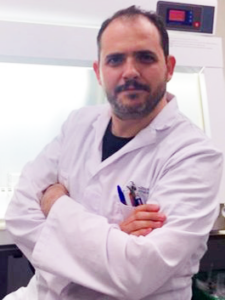 Ein wichtiger Forschungsbereich im Labor von Dr. Villa-Bellosta ist die übermäßige Verkalkung des Herzkreislaufsystems, einschließlich der Aorta, der Koronararterie und der Aortenklappen, die weitgehend die frühe Sterblichkeit bei Kindern mit HGPS bestimmt. Der molekulare Mechanismus der Gefäßverkalkung bei HGPS wurde zuvor an LmnaG609G/+ Knock-in-Mäusen analysiert, die einen starken Mangel an extrazellulärem Pyrophosphat aufweisen, einem wichtigen endogenen Inhibitor der Verkalkung. In diesem Projekt wollen wir die molekularen Mechanismen bestimmen, die die Gefäßverkalkung und die Langlebigkeit bei HGPS fördern oder verringern, wobei wir uns auf die Bedeutung bestimmter Nährstoffe konzentrieren, die täglich aufgenommen werden. Darüber hinaus planen wir, die Wirksamkeit von zwei neuen potenziellen therapeutischen Ansätzen (die die Pyrophosphathomöostase wiederherstellen) zu analysieren, die die Lebensqualität und Langlebigkeit von HGPS-Mäusen und Kindern verbessern könnten. Wir planen, LmnaG609G/+-Knock-in-Mäuse und glatte Gefäßmuskelzellen der Aorta zu verwenden, um die Wirkung dieser Nährstoffe/Behandlungen auf die Gefäßverkalkung und die Lebensdauer in vivo sowohl allein als auch in Kombination mit FTI-Lonafarnib zu analysieren.
November 2022: an Silvia Ortega Gutierrez, Universität Complutense, Madrid, Spanien
„Senkung des Progerinspiegels durch kleine Moleküle als neuer Ansatz zur Behandlung von Progerie“
Jüngste Erkenntnisse legen nahe, dass der wichtigste Einzelfaktor für den tödlichen Ausgang des Hutchinson-Gilford-Progerie-Syndroms (HGPS oder Progerie) die Ansammlung von Progerin ist, der mutierten Form von Lamin A, die Progerie verursacht. Genetische Ansätze, die darauf abzielen, die Progerinwerte entweder durch Interaktion mit seiner RNA oder durch Genkorrektur zu senken, führen zu erheblichen Verbesserungen des Krankheitsphänotyps. In diesem Projekt werden wir uns mit der direkten Reduzierung von Progerin durch die Entwicklung und Synthese kleiner Moleküle befassen, die als Proteolyse-Zielchimären (PROTACs) bezeichnet werden. Diese Klasse von Verbindungen, die hauptsächlich im letzten Jahrzehnt für andere Krankheiten entwickelt wurde, kann ein Protein spezifisch binden und es für den proteosomalen Abbau markieren und so seine Werte senken. Ausgehend von einem zuvor in unserem Labor identifizierten Treffer werden wir ein medizinalchemisches Programm durchführen, das darauf abzielt, verbesserte Verbindungen in Bezug auf biologische Aktivität und pharmakokinetische Parameter zu erhalten. Die optimale(n) Verbindung(en) werden auf ihre Wirksamkeit in einem In-vivo-Modell der Progerie untersucht.
 Oktober 2022: an Laurence Arbibe, Institut Necker-Enfants Malades (INEM), Paris, Frankreich
„Enthüllung der beschleunigten Darmalterung in der Pathophysiologie des HGPS: ein integrativer Ansatz“
Dr. Arbibes Labor zeigte kürzlich, dass chronische EntzündungenQualitätskontrolle des prä-mRNA-Spleißens im Darm, was unter anderem zur Produktion des Progerin-Proteins führt. Im vorliegenden Projekt wird sie die Auswirkungen der Progerin-Toxizität auf das Darmepithel untersuchen, Überwachung der Auswirkungen auf die Stammzellerneuerung und die Integrität der Schleimhautbarriere. Sie wird auch versuchen, pro-alternde Umweltreize zu identifizieren, die das RNA-Spleißen in HGPS beeinflussen, indem sie ein Reporter-Mausmodell implementiert, das in vivo Verfolgung des Progerin-spezifischen Spleißereignisses. Insgesamt befasst sich dieses Projekt mit den Auswirkungen der Progerie-Erkrankung auf die Integrität des Darms und stellt der wissenschaftlichen Gemeinschaft gleichzeitig neue Ressourcen für die Untersuchung gewebe- und zellspezifischer Ursachen für eine beschleunigte Alterung bei HGPS zur Verfügung.

Januar 2022: an Dr. Karima Djabali, PhD, Technische Universität München, München, Deutschland: „Behandlung des Hutchinson-Gilford-Progerie-Syndroms mit zwei von der FDA zugelassenen kombinierten Medikamenten — Lonafarnib Und Baricitinib, spezifische Inhibitoren der Farnesyltransferase bzw. der JAK1/2-Kinase.“
Dr. Djabalis Projekt wird in einem Mausmodell des HGPS testen, ob die Behandlung mit der Kombination von Lonafarnib Und Baricitinib, ein entzündungshemmendes Medikament, verzögert die Entwicklung der typischen HGPS-Pathologien, nämlich Gefäßerkrankungen, Hautatrophie, Alopezie und Lipodystrophie. Ihre früheren Erkenntnisse verknüpfen den JAK-STAT-Signalweg mit Entzündungen und zellulären Krankheitsmerkmalen von HGPS. Die zelluläre Exposition von HGPS gegenüber Baricitinib verbesserte das Zellwachstum und die mitochondriale Funktion, reduzierte entzündungsfördernde Faktoren, senkte den Progerinspiegel und verbesserte die Adipogenese. Darüber hinaus verbesserte die Verabreichung von Baricitinib mit Lonafarnib einige zelluläre Phänotypen über die alleinige Gabe von Lonafarnib hinaus.

Juli 2021: an Chiara Lanzuolo, Instituto Nazionale Genetica Molecolare, Mailand, Italien.
„Überwachung der Wiederherstellung der Genomstruktur und -funktion nach pharmakologischen Behandlungen beim Hutchinson-Gilford-Progerie-Syndrom“
Dr. Lanzuolo ist Expertin auf dem Gebiet der 3D-Struktur der DNA. Ihre Gruppe berichtete kürzlich, dass die zellspezifische dreidimensionale Struktur des Genoms durch die korrekte Anordnung der Kernlamina aufrechterhalten wird und bei der Pathogenese von Progerie schnell verloren geht. In diesem Projekt wird sie modernste Technologien an einem progerischen Mausmodell anwenden, um sich speziell mit den molekularen Mechanismen zu befassen, die in den frühen Phasen der Krankheit stattfinden und den Ausbruch der Krankheit entweder ermöglichen oder beschleunigen. Darüber hinaus wird sie die funktionelle Wiederherstellung des Genoms nach pharmakologischen Behandlungen analysieren.
 Juli 2021: an Mario Cordero, Institut für biomedizinische Forschung und Innovation von Cadiz (INIBICA), Cadiz, Spanien.
„Inflammasomhemmung und Polypillstrategie bei der Behandlung von HGPS“
Dr. Corderos Projekt wird die molekularen Auswirkungen des NLRP3-Inflammasom-Komplexes auf die Pathophysiologie von Progerie erforschen und die Auswirkungen eines spezifischen Inhibitors des NLRP3-Inflammasoms mit Lonafarnib untersuchen. Seine bisherigen Erkenntnisse zeigen eine mögliche Rolle von NLRP3 und eine mögliche Auswirkung seiner Hemmung auf das Überleben eines Progerie-Mausmodells. Er wird nun die Einzelmedikamentenbehandlung mit Lonafarnib mit einem spezifischen Inhibitor von NLRP3 und einer Kombinationsbehandlung aus beiden vergleichen, um festzustellen, welche am wirksamsten ist. Die Ergebnisse dieses Projekts werden hoffentlich dazu beitragen, eine klinische Studie zu Progerie mit zwei Verbindungen zu beschleunigen, die in Phase-2a-Studien am Menschen mit guter Wirkung und Verträglichkeit getestet wurden.

Juli 2020: (Startdatum August 2020) an Elsa Logarinho, Aging and Aneuploidy Group, IBMC – Instituto de Biologia Molecular e Celular, Porto, Portugal, „Steigerung der Chromosomenstabilität durch kleine Moleküle als senotherapeutische Strategie für HGPS“
Dr. Logarinhos Projekt zielt darauf ab, die Auswirkungen eines niedermolekularen Agonisten des Mikrotubuli (MT)-depolymerisierenden Kinesins-13 Kif2C/MCAK (UMK57) zu untersuchen, um den zellulären und physiologischen Merkmalen von HGPS entgegenzuwirken. Ihre bisherigen Erkenntnisse stufen Kif2C als Schlüsselfaktor sowohl bei genomischer als auch chromosomaler Instabilität ein, die ursächlich miteinander verbunden sind und auch als Hauptursache für Progerie-Syndrome gelten. Die Stabilisierung der Progerie-Chromosomen auf zellulärer Ebene zielt darauf ab, die Krankheit im gesamten Körper zu verbessern.
 Januar 2020: an Dr. Vicente Andrés, PhD, Centro Nacional de Investigaciones Cardioculares (CNIC), Madrid, Spanien. „Erzeugung transgener Lamin C-Stop (LCS) und CAG-Cre Yucatan-Minischweine zur Zucht von HGPS-Yucatan-Minischweinen für präklinische Versuche“
Ein Schwerpunkt der Forschung im Labor von Dr. Andrés ist die Entwicklung neuer Tiermodelle für Progeria. Große Tiermodelle bilden die Hauptmerkmale menschlicher Krankheiten viel besser nach als Mausmodelle, sodass wir Herz-Kreislauf-Erkrankungen untersuchen und Therapien testen können. Das Modell von Dr. Andrés wird ein neues Minischweinmodell für Progeria verbessern, das zuvor von PRF finanziert wurde.
 Januar 2020: an Dr. Giovanna Lattanzi, PhD, CNR Institute of Molecular Genetics Unit in Bologna, Italien. „Verbesserung der Lebensqualität bei Progerie: Ein erster Versuch im murinen LmnaG609G/G609G-Modell“
Dr. Lattanzi wird sich mit der Lebensqualität bei Progerie befassen, die mit einem chronischen Entzündungszustand verbunden ist. Die Normalisierung des Entzündungszustands kann Patienten helfen, pharmakologische Behandlungen zu überstehen; wenn sich ihr Gesundheitszustand verbessert, können sie eine bessere Wirksamkeit erzielen und ihre Lebensdauer verlängern. Dr. Lattanzi wird Strategien zur Reduzierung chronischer Entzündungen an einem Progerie-Mausmodell testen, mit dem Ziel, die Ergebnisse auf Patienten zu übertragen.
 Januar 2020: an Dr. Bum-Joon Park, PhD, Pusan National University, Republik Korea. „Wirkung von Progerinin (SLC-D011) und Lonafarnib auf HGPS: Eine kombinierte in vitro und in vivo“-Studie“
Dr. Park hat ein Medikament namens Progerinin entwickelt, das Progerin hemmt und die Krankheit in Progeriezellen bei Mäusen hemmt. Dr. Park wird nun die synergistischen Effekte von Progerinin mit Lonafarnib untersuchen. Er wird eine Einzelmedikamentenbehandlung (Lonafarnib) und eine Kombinationsbehandlung (Progerinin und Lonafarnib) vergleichen, um festzustellen, welche am wirksamsten ist. Wenn die Medikamentenkombination wenig toxisch ist, könnte eine kombinierte klinische Studie von Progerinin und Lonafarnib in Sicht sein!
 Januar 2020: An David R. Liu, PhD, Richard Merkin Professor und Direktor des Merkin Institute of Transformative Technologiesin Healthcare, Direktor des Chemical Biology and Therapeutic Sciences Program, Mitglied des Core Institute und stellvertretender Vorsitzender der Fakultät, Broad Institute, Forscher, Howard Hughes Medical Institute, Thomas Dudley Cabot Professor der Naturwissenschaften und Professor für Chemie und chemische Biologie, Harvard University. „Baseneditierungsbehandlungen für HGPS“.
Dr. Lius Labor wird Tests und Validierungen neuer Basen-Editor-Varianten durchführen, um das pathogene G608G-Allel zurück zum Wildtyp LMNA zu korrigieren, sowie die Entwicklung und Produktion von Viren, um diesen Editor und die entsprechende Leit-RNA in Patientenzellen zu bringen, die Entwicklung und Produktion der Viren, um diesen Editor und die entsprechende Leit-RNA in vivo zu bringen, Off-Target-DNA- und Off-Target-RNA-Analysen, RNA- und Proteinanalysen von behandelten Patientenzellen und Unterstützung für zusätzliche Experimente und Analysen, die benötigt werden, durchführen.
 Dezember 2019: Dr. Abigail Buchwalter ist Assistenzprofessorin am Cardiovascular Research Institute und der Abteilung für Physiologie der University of California in San Francisco. Die Projekte im Buchwalter-Labor konzentrieren sich auf die Definition der Mechanismen, die die Entstehung, Spezialisierung und Aufrechterhaltung der Kernorganisation über Zelltypen hinweg steuern. Von besonderem Interesse ist die Rolle der Kernlamina bei der Organisation des Genoms im Zellkern und die Definition, wie diese Ordnung durch krankheitsbedingte Mutationen gestört wird.
 Oktober 2019: Dr. Stewart ist ein sehr erfahrener Forscher auf dem Gebiet der Progerieforschung. Im letzten Jahrzehnt konzentrierte sich seine Forschung auf Laminopathien, eine heterogene Ansammlung von Krankheiten, die alle durch Mutationen im LaminA-Gen entstehen und sich auf Alterung, Herz-Kreislauf-Funktion und Muskeldystrophie auswirken. Er und seine Kollegen haben gezeigt, dass die Löschung eines Proteins namens SUN1 den Gewichtsverlust umkehrt und die Überlebensrate bei progerieähnlichen Mäusen erhöht. Auf der Grundlage dieser Erkenntnisse wird er nun ein Arzneimittelscreening durchführen und Tausende von Chemikalien auf solche untersuchen, die SUN1 stören und möglicherweise als neue Medikamente zur Behandlung von Kindern mit Progerie dienen könnten.
 November 2017: an Dr. Martin Bergö, PhD, Professor für Biowissenschaften, Karolinska Institute, Stockholm. „Entwicklung und präklinische Tests von ICMT-Inhibitoren für die HGPS-Therapie.“ Dr. Bergös Forschung basiert auf der Erkenntnis, dass die Reduktion von ICMT, einem Enzym, das für die Verarbeitung von Progerin benötigt wird, viele der pathologischen Merkmale bei Zmpste24-defizienten, progerieähnlichen Mäusen umkehrt. Seine vorläufigen Studien zeigen, dass im Labor gezüchtete Progeriezellen schneller und länger wachsen, wenn sie mit ICMT-Inhibitoren behandelt werden. Dr. Bergö wird Medikamente testen, die dieses Enzym blockieren und damit möglicherweise die Produktion von Progerin blockieren, und untersuchen, ob Progerie-Mausmodelle gesünder werden und länger leben, wenn sie mit dieser Art von Medikamenten behandelt werden.
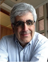 Arteriensteifigkeit bei HGPS: Auswirkungen auf die Lebensdauer.“ Dr. Assoian ist der Ansicht, dass ihre Forschung untersuchen wird, warum Arterien bei HGPS vorzeitig steif werden und ob vorzeitige Arteriensteifigkeit entweder durch pharmakologische Behandlung oder genetische Modifikation von Mäusen verhindert werden kann. Dr. Richard Assoian erhielt seine Ausbildung an der Johns Hopkins University (BA), der University of Chicago (PhD) und den National Institutes of Health (Postdoktorat). Er war an den Fakultäten der Columbia University und der University of Miami tätig, bevor er 1998 an die University of Pennsylvania wechselte. Derzeit ist er Professor für Pharmakologie in der Abteilung für Systempharmakologie und translationale Therapeutik der School of Medicine. Dr. Assoians Labor untersucht, wie Änderungen der Steifigkeit der arteriellen extrazellulären Matrix die Funktion der arteriellen glatten Muskelzellen beeinflussen. In dieser aktuellen Studie wird sein Labor ein Progeria-Mausmodell verwenden, um die Grundlagen und Folgen der vorzeitigen Arteriensteifigkeit bei HGPS zu untersuchen.
Dr. Finkel versucht zu verstehen, warum HGPS eine segmentale Progerie ist, also warum sie bestimmte Gewebe stärker zu beeinträchtigen scheint als andere. Er interessiert sich besonders dafür, warum Probleme mit Blutgefäßen auftreten. Man geht davon aus, dass diese segmentale Natur der Krankheit darauf zurückzuführen sein könnte, dass die Zelle, die zur Bildung der Blutgefäße beiträgt, die vaskuläre glatte Muskelzelle, möglicherweise etwas anders auf die Progerinexpression reagiert als andere Zelltypen. Dieser Unterschied hängt mit einem anderen Protein namens p62 zusammen, das am zellulären Prozess der Autophagie beteiligt ist. Er glaubt, dass sich p62 in glatten Muskelzellen anders verhält als in anderen Zellen (in glatten Muskelzellen scheint es sich im Zellkern anzusiedeln) und dass diese Unterschiede erklären könnten, warum die Blutgefäße bei HGPS so viele Probleme haben. Er glaubt auch, dass Medikamente entwickelt werden können, die auf p62 wirken, und dass diese Medikamente zur Behandlung von HGPS-Patienten nützlich sein könnten.
Toren Finkel ist Direktor des Aging Institute an der University of Pittsburgh/UPMC und Inhaber des G. Nicholas Beckwith III und Dorothy B. Beckwith Lehrstuhls für Translationale Medizin an der medizinischen Fakultät der University of Pittsburgh. Er erhielt 1986 seinen Bachelor-Abschluss in Physik sowie seinen MD- und PhD-Abschluss an der Harvard Medical School. Nach einer Facharztausbildung in Innerer Medizin am Massachusetts General Hospital absolvierte er ein Fellowship in Kardiologie an der Johns Hopkins Medical School. 1992 kam er als Forscher im Rahmen des Intramural Research Program des National Heart, Lung and Blood Institute (NHLBI) zum NIH. Während seiner Zeit am NIH hatte er verschiedene Positionen inne, darunter die des Leiters der Abteilung für Kardiologie und des Leiters des Zentrums für Molekulare Medizin am NHLBI. Er ist Mitglied der American Society for Clinical Research (ASCR), der Association of American Physicians (AAP) und Fellow der American Association for the Advancement of Science (AAAS). Er ist Mitglied zahlreicher Redaktionsausschüsse, darunter derzeit des Board of Reviewing Editors für Wissenschaft. Obwohl seine Arbeit hauptsächlich durch NIH Intramural Funds finanziert wurde, erhielt sein Labor Unterstützung als Senior Scholar der Ellison Medical Foundation und der Leducq Foundation, wo er derzeit als US-Koordinator für ein transatlantisches Netzwerk zur Erforschung der Herzregeneration fungiert. Seine aktuellen Forschungsinteressen umfassen die Rolle der Autophagie, reaktiver Sauerstoffspezies und der mitochondrialen Funktion bei der Alterung und altersbedingten Krankheiten.
 Herz-Kreislauf-Veränderungen sind die häufigste Todesursache bei Progerie-Patienten. Das Labor von Dr. Izpisua Belmonte hat nachgewiesen, dass durch zelluläre Neuprogrammierung Progerie-Zellen verjüngt werden können. Sein Labor verwendet nun zelluläre Neuprogrammierung, um Alterungsphänotypen in Mausmodellen von Progerie zu verbessern, wobei der Schwerpunkt auf dem Herz-Kreislauf-System liegt. Diese Entdeckungen könnten zur Entwicklung neuartiger Behandlungen für Progerie-Patienten führen.
Dr. Izpisua Belmontes Forschungsgebiet konzentriert sich auf das Verständnis der Stammzellbiologie sowie der Entwicklung und Regeneration von Organen und Geweben. Er hat über 350 Artikel in renommierten, international anerkannten, von Experten begutachteten Fachzeitschriften und Buchkapiteln veröffentlicht. Für seine Bemühungen auf diesen Gebieten erhielt er mehrere bemerkenswerte Ehrungen und Auszeichnungen, darunter den William Clinton Presidential Award, den Pew Scholar Award, den National Science Foundation Creativity Award, den American Heart Association Established Investigator Award und den Roger Guillemin Nobel Chair. Im Laufe der Jahre hat seine Arbeit dazu beigetragen, die Rolle einiger Homöobox-Gene bei der Strukturierung und Spezifikation von Organen und Geweben aufzudecken, sowie die molekularen Mechanismen zu identifizieren, die bestimmen, wie die verschiedenen Zelltypenvorläufer innerer Organe räumlich entlang der embryonalen Links-Rechts-Achse angeordnet sind. Seine Arbeit trägt dazu bei, uns einen Einblick in die molekularen Grundlagen zu geben, die bei der Organregeneration bei höheren Wirbeltieren, der Differenzierung menschlicher Stammzellen in verschiedene Gewebe sowie beim Altern und altersbedingten Krankheiten eine Rolle spielen. Das ultimative Ziel seiner Forschung ist die Entwicklung neuer Moleküle und spezifischer gen- und zellbasierter Behandlungsmethoden zur Heilung menschlicher Krankheiten.
Dezember 2016 (Startdatum 1. Februar 2017): An Ricardo Villa-Bellosta, PhD, Teamleiter, Fundación Jiménez Díaz University Hospital Health Research Institute (FIIS-FJD, Spanien). „Therapeutische Strategien zur Wiederherstellung der normalen Pyrophosphat-Homöostase bei HGPS.“
 Wie HGPS-Patienten, LmnaG609G/+ Mäuse weisen eine übermäßige Gefäßverkalkung auf, da die Fähigkeit des Körpers, extrazelluläres Pyrophosphat (PPi) zu synthetisieren, beeinträchtigt ist. Da ein Ungleichgewicht zwischen Abbau und Synthese von extrazellulärem PPi auch zu einer pathologischen Verkalkung von Gelenkknorpel und anderen Weichteilen führen kann, könnte der systemische Rückgang des zirkulierenden PPi, der mit der Progerin-Expression einhergeht, mehrere klinische Manifestationen von HGPS erklären, darunter Gefäßverkalkung sowie Knochen- und Gelenkanomalien. Die Behandlung mit exogenem PPi reduzierte die Gefäßverkalkung, verlängerte jedoch nicht die Lebensdauer von LmnaG609G/G609G Mäuse. Dies ist auf die schnelle Hydrolyse von exogenem PPi auf den basalen Serumspiegel zurückzuführen, wodurch die Wirkungszeit von PPi verkürzt wird, um ektopische Verkalkung in anderen Weichteilen wie Gelenken zu verhindern. Wiederherstellung der richtigen PPi-Homöostase in LmnaG609G/+Bei Mäusen, die pharmakologische Inhibitoren der am extrazellulären Pyrophosphat-Stoffwechsel beteiligten Enzyme verwenden, könnten sowohl die Lebensqualität als auch die Lebenserwartung verbessert werden.
Ricardo Villa-Bellosta promovierte 2010 an der Universität Saragossa (Spanien). Seine Doktorarbeit beschäftigte sich mit der Rolle von Phosphattransportern bei Gefäßverkalkung, Nierenphysiologie und Toxikokinetik von Arsen. Für seine Arbeit erhielt er mehrere Auszeichnungen, darunter den außerordentlichen Doktorandenpreis, den Preis der spanischen Königlichen Akademie der Ärzte und den Enrique Coris Research Award. Er war Gastforscher an der Emory University School of Medicine in Atlanta (USA), wo er den extrazellulären Pyrophosphat-Stoffwechsel (ePPi) in der Aortenwand untersuchte. 2012 wechselte er als Juan de la Cierva-Postdoktorand zum Centro Nacional de Investigaciones Cardiovasculares (CNIC, Spanien) und konzentrierte seine Arbeit auf den ePPi-Stoffwechsel sowohl bei der Verkalkung von Atheromplaques als auch bei der Gefäßverkalkung bei HGPS-Mäusen. 2015 wechselte er an das Fundación Jiménez Díaz University Hospital Health Research Institute (FIIS-FJD, Spanien), um dort als Postdoktorand bei Sara Borrell die Phosphat-/Pyrophosphat-Homöostase bei Hämodialysepatienten zu untersuchen. Im September 2015 erhielt er als Teamleiter am FIIS-FJD ein „I+D+I Young Researchers“-Stipendium, um die Rolle des ePPi-Stoffwechsels bei der Gefäßverkalkung bei chronischer Nierenerkrankung und Diabetes zu untersuchen.
 Die ursächliche Mutation von HGPS betrifft Lamin A. AKTIP, ein Protein, das wir kürzlich charakterisiert haben, ist ein mit Lamin interagierender Faktor, der für das Zellüberleben unerlässlich ist und am Telomer- und DNA-Stoffwechsel beteiligt ist. Vier Hauptbeobachtungen bringen dieses neue Protein mit HGPS in Verbindung: i) AKTIP-Beeinträchtigung rekapituliert HGPS-Eigenschaften in Zellen; ii) AKTIP-Beeinträchtigung rekapituliert HGPS-Eigenschaften in Mäusen; iii) AKTIP interagiert mit Laminen und iv) AKTIP ist in von Patienten stammenden HGPS-Zellen verändert. In unseren Studien postulieren wir die Hypothese, dass ein AKTIP-Komplex als Kontrollpunkt für herausfordernde DNA-Replikationsereignisse fungiert. Wir erwarten, dass dieser Kontrollpunkt bei HGPS beeinträchtigt ist, was wiederum zum HGPS-Phänotyp beitragen kann. Wir schlagen vor, die AKTIP-Funktion in vitro und bei Mäusen umfassend zu analysieren. Wir erwarten, dass diese Forschung neue Einblicke in den Zusammenhang zwischen Progerin und Telomerfunktionsstörungen durch AKTIP sowie Informationen über die Rolle der DNA-Replikationsstörung als potenzieller Treibermechanismus bei Progerie liefert. Da das Wissen über die Determinanten und Treibermechanismen der HGPS-Ätiologie noch nicht vollständig ist, glauben wir, dass Studien zu neuen mit Lamin interagierenden Akteuren wie AKTIP von entscheidender Bedeutung sein werden, um die mechanistischen Grundlagen von HGPS zu entschlüsseln und den Weg für neue therapeutische Strategien zu ebnen.
Isabella Saggio erhielt ihren Doktortitel in Genetik an der Sapienza-Universität (Rom, Italien). Von 1991 bis 1994 arbeitete sie am Merck Research Institute for Molecular Biology (Rom, Italien). Von 1994 bis 1997 war sie EU-Postdoktorandin am IGR (Paris, Frankreich). 1998 kehrte sie an die Sapienza-Universität zurück, zunächst als wissenschaftliche Mitarbeiterin und dann als außerordentliche Professorin für Genetik und Gentherapie. IS Hauptforschungsinteressen sind Gentherapie sowie Studien zu Telomeren und Alterung. IS war von 2003 bis 2011 Mitglied des San Raffaele Science Park, ist seit 2003 Teil des CNR und seit 2016 des italienischen Netzwerks für Laminopathien. IS ist die Sapienza-Vertreterin im Interuniversity Biotechnology Network in Italien, koordiniert internationale Aktivitäten an der Sapienza und gründete 2016 einen Master of Scientific Journalism, um die Beziehungen zwischen Forschern und der Öffentlichkeit zu verbessern (www.mastersgp.it). IS-Aktivitäten werden auf der Site beschrieben: www.saggiolab.com.
.jpg) Unser Ziel ist es, neue potenzielle Progerie-Therapeutika in vivo zu testen. Dieses stark kollaborative Projekt basiert auf der Entdeckung mehrerer potenzieller Therapeutika im Labor von Tom Misteli, der Entwicklung eines HGPS-Tiermodells im Labor von Carlos Lopez-Otin und der Expertise von Alicia Rodriguez-Folgueras beim Testen verschiedener Verbindungen in einer In-vivo-Umgebung.
Unser Ziel ist es, neue potenzielle Progerie-Therapeutika in vivo zu testen. Dieses stark kollaborative Projekt basiert auf der Entdeckung mehrerer potenzieller Therapeutika im Labor von Tom Misteli, der Entwicklung eines HGPS-Tiermodells im Labor von Carlos Lopez-Otin und der Expertise von Alicia Rodriguez-Folgueras beim Testen verschiedener Verbindungen in einer In-vivo-Umgebung.
Tom Misteli ist ein NIH Distinguished Investigator und Direktor des Center for Cancer Research am National Cancer Institute, NIH. Er ist ein international renommierter Zellbiologe, der Pionierarbeit bei der Verwendung von Bildgebungsverfahren zur Untersuchung von Genomen und Genexpression in lebenden Zellen geleistet hat. Das Interesse seines Labors besteht darin, die grundlegenden Prinzipien der dreidimensionalen Genomorganisation und -funktion aufzudecken und dieses Wissen für die Entwicklung neuer diagnostischer und therapeutischer Strategien für Krebs und Alterung anzuwenden. Er erhielt seinen Doktortitel an der University of London, Großbritannien, und absolvierte eine Postdoc-Ausbildung am Cold Spring Harbor Laboratory. Für seine Arbeit erhielt er zahlreiche Auszeichnungen, darunter den Herman Beerman Award, die Wilhelm Bernhard Medaille, die Goldmedaille der Karls-Universität, den Flemming Award, den Gian-Tondury-Preis, den NIH Director's Award und einen NIH Merit Award. Er fungiert als Berater für zahlreiche nationale und internationale Agenturen und ist Mitglied mehrerer Redaktionsausschüsse, darunter Zelle, Wissenschaft Und PLoS-Biologie. Er ist der Chefredakteur von Aktuelle Meinung in der Zellbiologie.
 In diesem Projekt schlagen wir die Entwicklung neuer Inhibitoren der Isoprenylcystein-Carboxylmethyltransferase (ICMT) zur Behandlung des Hutchinson-Gilford-Progerie-Syndroms (HGPS oder Progerie) vor, basierend auf einem zuvor in unserem Forschungslabor identifizierten Hit. Dieser Hit (UCM-13239) hemmt ICMT in signifikanter Weise, induziert eine Fehllokalisierung des Progerinproteins in progeroiden Fibroblasten (LmnaG609G/G609G), erhöht die Lebensfähigkeit dieser Zellen und fördert überlebensfördernde Signalwege in den behandelten Zellen. Mit dieser Verbindung als Ausgangspunkt wird unser Team ein medizinalchemisches Programm (Hit-to-Lead und Lead-Optimierung) durchführen, das darauf abzielt, verbesserte Verbindungen in Bezug auf biologische Aktivität und pharmakokinetische Parameter zu erhalten. Die Wirksamkeit der optimalen Verbindung(en) wird in einem In-vivo-Modell der Progerie bewertet.
Silvia Ortega-Gutiérrez promovierte an der Universität Complutense in Madrid unter der Aufsicht von Prof. María Luz López-Rodríguez im Bereich der medizinischen Chemie. Danach wechselte sie mit einem Fulbright-Stipendium in das Labor von Prof. Ben Cravatt am Scripps Research Institute (Kalifornien, USA), um im Bereich der chemischen Biologie und Proteomik zu arbeiten. Zwischen 2008 und 2012 war sie Ramón y Cajal-Stipendiatin in der Abteilung für organische Chemie der Universität Complutense, wo sie 2013 zur außerordentlichen Professorin befördert wurde. Diese Position hat sie derzeit inne.
Die Interessengebiete von Dr. Ortega-Gutiérrez sind die medizinische Chemie und die chemische Biologie, insbesondere die Bereiche der endogenen Cannabinoid- und Lysophosphatidsäuresysteme, die Validierung neuer therapeutischer Ziele und die Entwicklung chemischer Sonden für die Untersuchung von G-Protein-gekoppelten Rezeptoren. Ihre Arbeiten wurden in renommierten Zeitschriften wie Science, Nature Neuroscience, Angewandte Chemie und dem Journal of Medicinal Chemistry veröffentlicht und auch in Patenten, die an die Pharmaindustrie übertragen wurden. 2011 und 2016 erhielt sie den „Runner-up Prize for a Young Medicinal Chemist in Academia“ der European Federation of Medicinal Chemistry und 2012 den „Young Researcher Award“ der spanischen Royal Chemical Society.
 Das Hutchinson-Gilford-Progerie-Syndrom (HGPS) ist eine seltene genetische Erkrankung, die durch eine Mutation im LMNA Gen und gekennzeichnet durch schwere Symptome, die Merkmalen vorzeitiger Alterung ähneln, einschließlich Herz-Kreislauf-Erkrankungen, die zu Arteriosklerose, Bluthochdruck, Herzhypertrophie und Tod durch Herzversagen führen. Frühere Studien an Patienten und HGPS-Mausmodellen zeigten einen fortschreitenden Verlust von glatten Muskelzellen in Blutgefäßen, aber die Rolle von Endothelzellen bei der Entwicklung von HGPS-bedingten Herz-Kreislauf-Erkrankungen wurde noch nicht analysiert, obwohl die beeinträchtigte Funktion der Endothelzellen ein Hauptrisikofaktor für Herz-Kreislauf-Erkrankungen bei normalem Altern ist. Um die molekulare Basis der kardiovaskulären Alterungspathologie zu untersuchen und zu erforschen, wie das gealterte Gefäßendothel zu HGPS beiträgt, haben wir ein neuartiges Mausmodell entwickelt, das das HGPS-verursachende LMNA mutiertes Genprodukt selektiv im vaskulären Endothelzellsystem. Unsere vorläufigen Analysen der Mäuse zeigten verzögertes Wachstum, erhöhte Fibrose im Herzen, Herzhypertrophie, Erhöhung der Hypertrophiemarker und vorzeitigen Tod der mutierten Mäuse, was dem kardiovaskulären Phänotyp des HGPS ähnelt. In diesem Projekt werden wir die molekularen Mechanismen untersuchen, wie das mutierte LMNA Genprodukt beeinflusst Endothelzellen in Blutgefäßen und wie sich dies auf die Herzfunktion auswirken kann. Wir werden proatherogene Komponenten identifizieren, die in mutierten Endothelzellen und Gefäßen abgesondert werden, und testen, wie dieser Weg andere Gewebe und Zellen beeinflussen kann. Dieses Projekt wird auch potenzielle Biomarker für mit HGPS verbundene Herz-Kreislauf-Erkrankungen im Blut identifizieren. Unser Projekt untersucht zum ersten Mal die Rolle des Gefäßendothels bei der Entwicklung von Herz-Kreislauf-Erkrankungen bei HGPS und wird neue (proatherogene) Wege und Komponenten als potenzielle Ziele für Diagnose und Therapie identifizieren.
Roland Foisner ist Universitätsprofessor für Biochemie an der Medizinischen Universität Wien und stellvertretender Direktor der Max F. Perutz Laboratories. Er promovierte 1984 in Biotechnologie an der Technischen Universität Wien, Österreich, war Assistenz- und dann außerordentlicher Professor an der Universität Wien und wurde 2002 zum ordentlichen Professor an der Abteilung für Medizinische Biochemie der Medizinischen Universität Wien ernannt. 1991–1992 absolvierte er eine Postdoktorandenausbildung am Scripps Research Institute in La Jolla, Kalifornien, USA.
Roland Foisner war wissenschaftlicher Koordinator von EURO‐Laminopathies, einem europäischen Netzwerkprojekt klinischer und grundlagenorientierter Forscher, dessen Ziel die Analyse der molekularen Mechanismen von Laminopathien zur Entwicklung neuer Therapieansätze ist. Er ist Chefredakteur des Journals Nucleus, Mitglied des Redaktionsausschusses mehrerer zellbiologischer Journale, des wissenschaftlichen Beirats von EU‐Projekten und von Gutachtergremien mehrerer internationaler Förderorganisationen. Er war bis 2007 Dekan für Graduiertenstudien im PhD‐Programm des International Vienna Biocenter und war Mitglied zahlreicher nationaler und internationaler Promotionsausschüsse.
Die Forschung im Labor von Roland Foisner konzentriert sich auf die Dynamik und Funktionen von Laminen und Laminbindungsproteinen in der Kern- und Chromatinorganisation, in der Regulierung der Genexpression und Signalgebung sowie in genetischen Erkrankungen von Muskeldystrophien bis hin zu vorzeitiger Alterung. Er hat zahlreiche wichtige, von Experten begutachtete Artikel, eingeladene Rezensionen und Buchkapitel veröffentlicht und zahlreiche eingeladene Seminare bei nationalen und internationalen Tagungen gehalten.
 Herz-Kreislauf-Veränderungen sind die häufigste Todesursache bei Progerie-Patienten. Dr. Belmontes Labor hat neue Modelle für die Erforschung von Progerie entwickelt, die auf der Verwendung von induzierten pluripotenten Stammzellen (iPSCs) basieren, die von Progerie-Patienten erzeugt wurden. Sein Labor verwendet nun aus diesen Modellen erzeugte Gefäßzellen für die Entwicklung neuer Medikamente, die Herz-Kreislauf-Veränderungen in menschlichen und Mausmodellen von Progerie lindern können. Diese Entdeckungen könnten zur Entwicklung neuer Behandlungen für Progerie-Patienten führen.
Dr. Juan Carlos Belmonte Izpisua ist Professor an den Gene Expression Laboratories der Salk-Institut für biologische Studien, La Jolla, CA, USA. Er ist der ehemalige Direktor und half beim Aufbau der Zentrum für Regenerative Medizin in Barcelona. Er hat einen Doktortitel in Biochemie und Pharmakologie von der Universität Bologna, Italien und der Universität Valencia, Spanien. Er ist Postdoktorand am Europäischen Laboratorium für Molekularbiologie (EMBL) der Universität Marburg in Heidelberg, Deutschland, und an der UCLA, USA.
 Eine aktuelle Studie anderer Forscher [Gabriel et al., 2015, Alternde Zelle 14(1):78-91] zeigte, dass das Isothiocyanat Sulforaphan (ein Phytochemikal aus Brokkoli) die Wachstumsrate von kultivierten Zellen von Kindern mit Progerie steigerte und eine Reihe von Biomarkern erhöhte, die mit dem Syndrom in Zusammenhang stehen. Unsere Arbeit mit Isothiocyanaten aus essbaren Pflanzen legt nahe, dass einige dieser über hundert eng verwandten Verbindungen ein breiteres therapeutisches Fenster (den Bereich zwischen wirksamer und toxischer Konzentration) und möglicherweise niedrigere wirksame Konzentrationen als Sulforaphan haben sollten. Wir werden diese Hypothese testen.
.jpg) Wir haben kürzlich durch Screening einer chemischen Bibliothek neue Chemikalien gefunden, die die Interaktion zwischen Progerin und Lamin A/C blockieren. In einem Progerin-produzierenden Mausmodell (LmnaG609G/G609G), kann unsere Chemikalie (JH4) die Lebensdauer verlängern sowie Alterungserscheinungen wie Gewichtszunahme, Muskelkraft und Organgröße verbessern. Trotz der offensichtlichen Wirkung von JH4 auf Lmnawt/G609GMäuse, kann es nur 4 Wochen verlängern LmnaG609G/G609G Die Lebensspanne der Mäuse deutet darauf hin, dass die Wirkung von JH4 zum gegenwärtigen Zeitpunkt nicht ausreicht, um es als Therapeutikum für das Progerie-Syndrom einzusetzen. Darüber hinaus muss die Wirkung von JH4 verbessert werden. Dazu werden wir mehrere Versuche zur Verbesserung der Wirkung von JH4 durchführen. Zunächst werden wir unsere Chemikalien in eine hydrophilere Form bringen. Tatsächlich ist JH4 sehr hydrophob, was einer der Gründe ist, warum wir die Dosierung nicht erhöhen können. In dieser Hinsicht haben wir bereits eine hydrophile Verbindung (JH010) mit ähnlicher zellulärer Wirkung wie JH4 erhalten. Tatsächlich haben unsere jüngsten Ergebnisse gezeigt, dass eine Erhöhung von JH4 (von 10 mg/kg auf 20 mg/kg) die Lebensspanne von 16 Wochen (mit Träger behandelt) auf 24 Wochen erhöhen kann (tatsächlich waren Mäuse, denen 20 mg/kg injiziert wurden, noch am Leben). Um diese Chemikalie zu verbessern, haben wir JH010-Derivate erzeugt und die biologische Wirkung überprüft. Zweitens werden wir Nanopartikel herstellen, die JH010 effektiver an den gesamten Körper abgeben. Tatsächlich haben wir mit dieser Arbeit bereits begonnen. Durch beide Methoden werden wir verbesserte JH4-verwandte Chemikalien erhalten und diese testen in LmnaG609G/G609G Mausmodell (Lebensdauer, histologische Analyse, Toxizität, Pharmakodynamik sowie Pharmakokinetik). Auf Grundlage dieser Studien möchten wir die beste Behandlungsmethode für HGPS im Mausmodell sowie bei HGPS-Kindern entwickeln.
Wir haben kürzlich durch Screening einer chemischen Bibliothek neue Chemikalien gefunden, die die Interaktion zwischen Progerin und Lamin A/C blockieren. In einem Progerin-produzierenden Mausmodell (LmnaG609G/G609G), kann unsere Chemikalie (JH4) die Lebensdauer verlängern sowie Alterungserscheinungen wie Gewichtszunahme, Muskelkraft und Organgröße verbessern. Trotz der offensichtlichen Wirkung von JH4 auf Lmnawt/G609GMäuse, kann es nur 4 Wochen verlängern LmnaG609G/G609G Die Lebensspanne der Mäuse deutet darauf hin, dass die Wirkung von JH4 zum gegenwärtigen Zeitpunkt nicht ausreicht, um es als Therapeutikum für das Progerie-Syndrom einzusetzen. Darüber hinaus muss die Wirkung von JH4 verbessert werden. Dazu werden wir mehrere Versuche zur Verbesserung der Wirkung von JH4 durchführen. Zunächst werden wir unsere Chemikalien in eine hydrophilere Form bringen. Tatsächlich ist JH4 sehr hydrophob, was einer der Gründe ist, warum wir die Dosierung nicht erhöhen können. In dieser Hinsicht haben wir bereits eine hydrophile Verbindung (JH010) mit ähnlicher zellulärer Wirkung wie JH4 erhalten. Tatsächlich haben unsere jüngsten Ergebnisse gezeigt, dass eine Erhöhung von JH4 (von 10 mg/kg auf 20 mg/kg) die Lebensspanne von 16 Wochen (mit Träger behandelt) auf 24 Wochen erhöhen kann (tatsächlich waren Mäuse, denen 20 mg/kg injiziert wurden, noch am Leben). Um diese Chemikalie zu verbessern, haben wir JH010-Derivate erzeugt und die biologische Wirkung überprüft. Zweitens werden wir Nanopartikel herstellen, die JH010 effektiver an den gesamten Körper abgeben. Tatsächlich haben wir mit dieser Arbeit bereits begonnen. Durch beide Methoden werden wir verbesserte JH4-verwandte Chemikalien erhalten und diese testen in LmnaG609G/G609G Mausmodell (Lebensdauer, histologische Analyse, Toxizität, Pharmakodynamik sowie Pharmakokinetik). Auf Grundlage dieser Studien möchten wir die beste Behandlungsmethode für HGPS im Mausmodell sowie bei HGPS-Kindern entwickeln.
Dr. Park promovierte in Krebsbiologie an der Korea University. Nach seiner Promotion führte er seine Forschungsarbeiten am Korea National Institute of Health (KNIH) und an der Seoul National University durch. Seit 2006 arbeitet er an der Pusan National University. Derzeit ist er Vorsitzender der Abteilung für Molekularbiologie. Seine Forschung konzentriert sich auf die Identifizierung des krankheitsspezifischen Signalnetzwerks (Krebs, HGPS, Werner-Syndrom) und die Suche nach neuen Chemikalien, die die krankheitsbedingte Protein-Protein-Interaktion für Arzneimittelkandidaten blockieren können.
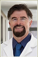 Bei Kindern mit Progerie altern die Blutgefäße sehr schnell. Dies führt zu Gefäßerkrankungen, die zu Herzinfarkten und Schlaganfällen führen. Wir beabsichtigen, eine Therapie zu entwickeln, die die Gefäßalterung bei diesen Kindern umkehrt. Wir haben bereits gezeigt, dass gealterte menschliche Zellen verjüngt werden können, indem man sie mit modifizierter Message-RNA (mmRNA) behandelt, die Telomerase kodiert. Telomerase ist ein Protein, das Telomere auf Chromosomen verlängert.
Die Telomere sind wie die Spitze eines Schnürsenkels; sie halten die Chromosomen zusammen und sind für die normale Funktion der Chromosomen notwendig. Wenn Zellen altern, werden die Telomere kürzer und irgendwann funktioniert das Chromosom nicht mehr richtig. An diesem Punkt altert die Zelle und kann sich nicht mehr vermehren. Telomere sind im Wesentlichen unsere biologische Uhr. Bei Kindern mit Progerie verkürzen sich die Telomere schneller. Wir beabsichtigen, unsere Therapie an Zellen von Progerie-Kindern zu testen, um zu sehen, ob wir die Telomere verlängern, den Alterungsprozess umkehren und die Gefäßzellen verjüngen können. Wenn dieser Ansatz funktioniert, beabsichtigen wir, die Therapie für klinische Studien an diesen Kindern weiterzuentwickeln.
Dr. John P. Cooke studierte Herz-Kreislaufmedizin und promovierte in Physiologie an der Mayo Clinic. Er wurde als Assistenzprofessor für Medizin an die Harvard Medical School berufen. 1990 wurde er an die Stanford University berufen, um dort das Programm für Gefäßbiologie und -medizin zu leiten. Er wurde zum Professor in der Abteilung für Herz-Kreislaufmedizin an der Stanford University School of Medicine und zum stellvertretenden Direktor des Stanford Cardiovascular Institute ernannt, bis er 2013 an die Houston Methodist berufen wurde.
Dr. Cooke hat über 500 Forschungsarbeiten, Positionspapiere, Rezensionen, Buchkapitel und Patente im Bereich der Gefäßmedizin und -biologie mit über 20.000 Zitierungen veröffentlicht; h-Index = 76 (ISI Web of Knowledge, 6.2.13). Er ist Mitglied nationaler und internationaler Ausschüsse, die sich mit Herz-Kreislauf-Erkrankungen befassen, darunter der American Heart Association, dem American College of Cardiology, der Society for Vascular Medicine und dem National Heart, Lung and Blood Institute. Er war Präsident der Society for Vascular Medicine, Direktor des American Board of Vascular Medicine und Mitherausgeber von Vascular Medicine.
Dr. Cookes translationales Forschungsprogramm konzentriert sich auf die Gefäßregeneration. Das Programm wird durch Zuschüsse der National Institutes of Health, der American Heart Association und der Industrie finanziert.
Der Schwerpunkt von Dr. Cookes Forschungsprogramm liegt auf der Wiederherstellung oder Stimulation von Endothelfunktionen wie Vasodilatation und Angiogenese durch die Verwendung kleiner Moleküle oder Stammzellentherapien. In seinen 25 Jahren translationaler Endothelbiologie beschrieb und charakterisierte er als Erster die antiatherogenen Effekte von aus Endothel gewonnenem Stickstoffmonoxid, die antiangiogene Wirkung des NO-Synthase-Hemmers ADMA, den durch endotheliale nikotinbedingte Acetylcholinrezeptoren vermittelten Angiogeneseweg, die Rolle dieses Weges in Zuständen pathologischer Angiogenese und entwickelte einen Antagonisten des Weges, der sich derzeit in Phase II klinischer Tests befindet. Seine klinische Forschungsgruppe untersuchte den Einsatz angiogener Wirkstoffe und adulter Stammzellen bei der Behandlung peripherer arterieller Verschlusskrankheit. Vor kurzem erzeugte und charakterisierte er aus menschlichen iPSCs gewonnene Endothelzellen und untersuchte ihre Rolle bei der Angiogenese und Gefäßregeneration. Aktuelle Erkenntnisse aus dem Labor haben die Rolle der angeborenen Immunsignale bei der nukleären Reprogrammierung zur Pluripotenz und der therapeutischen Transdifferenzierung bei Gefäßerkrankungen geklärt.
.jpg) Dr. Collins beaufsichtigt die Arbeit des weltweit größten Förderers der biomedizinischen Forschung, von der Grundlagenforschung bis zur klinischen Forschung. Dr. Collins und sein Team haben 2003 gemeinsam mit der Progeria Research Foundation die genetische Ursache von HGPS entdeckt, und nach über zwölf Jahren Arbeit bleibt ihr Ziel: die Pathogenese zu verstehen und Behandlungsmöglichkeiten für HGPS zu finden. Aktuelle Studien konzentrieren sich auf potenzielle therapeutische Ansätze, darunter RNA-basierte Methoden und die Verwendung von Rapamycin und seinen Analoga, wobei sowohl zelluläre als auch HGPS-Mausmodelle zum Einsatz kommen.
Dr. Collins beaufsichtigt die Arbeit des weltweit größten Förderers der biomedizinischen Forschung, von der Grundlagenforschung bis zur klinischen Forschung. Dr. Collins und sein Team haben 2003 gemeinsam mit der Progeria Research Foundation die genetische Ursache von HGPS entdeckt, und nach über zwölf Jahren Arbeit bleibt ihr Ziel: die Pathogenese zu verstehen und Behandlungsmöglichkeiten für HGPS zu finden. Aktuelle Studien konzentrieren sich auf potenzielle therapeutische Ansätze, darunter RNA-basierte Methoden und die Verwendung von Rapamycin und seinen Analoga, wobei sowohl zelluläre als auch HGPS-Mausmodelle zum Einsatz kommen.
Francis S. Collins, MD, Ph.D. ist Direktor der National Institutes of Health (NIH). In dieser Funktion beaufsichtigt er die Arbeit des größten Förderers der biomedizinischen Forschung weltweit, die das gesamte Spektrum von der Grundlagenforschung bis zur klinischen Forschung abdeckt.
Dr. Collins ist ein Arzt und Genetiker, der für seine bahnbrechenden Entdeckungen von Krankheitsgenen und seine Leitung des internationalen Humangenomprojekts bekannt ist, das im April 2003 mit der Fertigstellung einer vollständigen Sequenz der Anleitung zur menschlichen DNA seinen Höhepunkt erreichte. Von 1993 bis 2008 war er Direktor des National Human Genome Research Institute am NIH.
Dr. Collins‘ eigenes Forschungslabor hat eine Reihe wichtiger Gene entdeckt, darunter solche, die für Mukoviszidose, Neurofibromatose, die Huntington-Krankheit, ein familiäres endokrines Krebssyndrom und in jüngster Zeit Gene für Typ-2-Diabetes verantwortlich sind, sowie das Gen, das das Hutchinson-Gilford-Progerie-Syndrom verursacht, eine seltene Erkrankung, die vorzeitige Alterung zur Folge hat.
Dr. Collins erhielt einen BS in Chemie von der University of Virginia, einen Ph.D. in physikalischer Chemie von der Yale University und einen MD mit Auszeichnung von der University of North Carolina in Chapel Hill. Bevor er 1993 zum NIH kam, war er neun Jahre lang an der University of Michigan tätig, wo er als Forscher am Howard Hughes Medical Institute tätig war. Er ist gewähltes Mitglied des Institute of Medicine und der National Academy of Sciences. Dr. Collins wurde im November 2007 mit der Presidential Medal of Freedom und 2009 mit der National Medal of Science ausgezeichnet.
 Das Hutchinson-Gilford-Progerie-Syndrom (HGPS) ist eine seltene, tödliche genetische Störung, die durch schnelles Altern gekennzeichnet ist. Die Behandlung menschlicher HGPS-Fibroblasten oder Mäuse ohne Lmna (ein Mausmodell von HGPS) mit Rapamycin, einem Inhibitor der mTOR-Proteinkinase (mechanistic Target Of Rapamycin), kehrt HGPS-Phänotypen auf zellulärer Ebene um und fördert Lebensdauer und Gesundheit auf Organismusebene. Rapamycin hat jedoch schwerwiegende Nebenwirkungen beim Menschen, darunter Immunsuppression und diabetogene Stoffwechseleffekte, die seine langfristige Anwendung bei HGPS-Patienten ausschließen können. Die mTOR-Proteinkinase kommt in zwei unterschiedlichen Komplexen vor. Die Arbeit des Forschungsteams von Dr. Lamming sowie die Arbeit vieler anderer Labore legen nahe, dass viele der Vorteile von Rapamycin auf die Unterdrückung des mTOR-Komplexes 1 (mTORC1) zurückzuführen sind, während viele der Nebenwirkungen auf eine unerwünschte Hemmung des mTOR-Komplexes 2 (mTORC2) zurückzuführen sind.
Während Rapamycin beide mTOR-Komplexe in vivo hemmt, reagieren mTORC1 und mTORC2 natürlich auf verschiedene Umwelt- und Nährstoffreize. mTORC1 wird direkt durch Aminosäuren stimuliert, während mTORC2 hauptsächlich durch Insulin und Wachstumsfaktorsignale reguliert wird. Dr. Lammings Forschungsteam hat festgestellt, dass eine proteinarme Ernährung die mTORC1-, aber nicht die mTORC2-Signalgebung in Mäusegeweben signifikant reduziert. Dies wirft die faszinierende Möglichkeit auf, dass eine proteinarme Ernährung eine relativ einfache Methode mit geringen Nebenwirkungen sein könnte, um die mTORC1-Aktivität einzuschränken und HGPS-Patienten einen therapeutischen Nutzen zu bieten. In dieser Studie werden sie eine Ernährung identifizieren, die die mTORC1-Signalgebung in vivo hemmt, und die Fähigkeit dieser Ernährung bestimmen, die HGPS-Pathologie sowohl in vivo in einem Progerin-exprimierenden Mausmodell von HGPS als auch in vitro in menschlichen HGPS-Patientenzelllinien zu retten.
Dudley Lamming erhielt 2008 seinen Doktortitel in experimenteller Pathologie an der Harvard University im Labor von Dr. David Sinclair und absolvierte anschließend eine Postdoc-Ausbildung am Whitehead Institute for Biomedical Research in Cambridge, MA, im Labor von Dr. David Sabatini. Dr. Lammings Forschung wird teilweise durch einen NIH/NIA K99/R00 Pathway to Independence Award sowie einen Junior Faculty Research Award der American Federation for Aging Research unterstützt. Sein Labor an der University of Wisconsin konzentriert sich darauf, herauszufinden, wie nährstoffabhängige Signalwege genutzt werden können, um die Gesundheit zu fördern und sowohl normales Altern als auch vorzeitige Alterungskrankheiten wie das Hutchinson-Gilford-Progerie-Syndrom zu verzögern.
.jpg) Das Hutchinson-Gilford-Progerie-Syndrom (HGPS) ist eine äußerst seltene genetische Erkrankung, die durch vorzeitiges und beschleunigtes Altern und vorzeitigen Tod gekennzeichnet ist. Die Entdeckung neuer therapeutischer Verbindungen ist für diese tödliche Krankheit von größter Bedeutung. Das endogene Molekül Neuropeptid Y (NPY) aktiviert NPY-Rezeptoren, die in verschiedenen von HGPS betroffenen Organen und Zellen lokalisiert sind. Unsere vorläufigen Daten und jüngsten Veröffentlichungen legen stark nahe, dass das Neuropeptid Y (NPY)-System ein mutmaßliches therapeutisches Ziel für HGPS sein könnte.
Das Hutchinson-Gilford-Progerie-Syndrom (HGPS) ist eine äußerst seltene genetische Erkrankung, die durch vorzeitiges und beschleunigtes Altern und vorzeitigen Tod gekennzeichnet ist. Die Entdeckung neuer therapeutischer Verbindungen ist für diese tödliche Krankheit von größter Bedeutung. Das endogene Molekül Neuropeptid Y (NPY) aktiviert NPY-Rezeptoren, die in verschiedenen von HGPS betroffenen Organen und Zellen lokalisiert sind. Unsere vorläufigen Daten und jüngsten Veröffentlichungen legen stark nahe, dass das Neuropeptid Y (NPY)-System ein mutmaßliches therapeutisches Ziel für HGPS sein könnte.
In dieser Studie werden wir die positiven Auswirkungen von NPY und/oder Aktivatoren von NPY-Rezeptoren bei der Rettung des Alterungsphänotyps in zwei HGPS-Modellen untersuchen: in einem zellbasierten und einem Mausmodell von HGPS. Mit diesem Projekt wollen wir zeigen, dass die Aktivierung des NPY-Systems eine innovative Strategie für die Therapie oder Co-Therapie von HGPS ist.
Cláudia Cavadas hat einen Doktortitel in Pharmakologie von der Fakultät für Pharmazie der Universität Coimbra. Sie ist Gruppenleiterin der „Neuroendokrinologie und Alternsgruppe“ am CNC – Zentrum für Neurowissenschaften und Zellbiologie der Universität Coimbra. Cláudia Cavadas ist Mitautorin von 50 Veröffentlichungen und erforscht seit 1998 das Neuropeptid-Y-System (NPY). Sie ist Vizepräsidentin der portugiesischen Gesellschaft für Pharmakologie (seit 2013); Cláudia Cavadas war die ehemalige Direktorin des Instituts für interdisziplinäre Forschung der Universität Coimbra (2010-2012).
 Das Hutchinson-Gilford-Progerie-Syndrom (HGPS), eine tödliche genetische Störung, ist durch vorzeitiges, beschleunigtes Altern gekennzeichnet. HGPS wird am häufigsten durch eine De-novo-Punktmutation (G608G) im Lamin A/C-Gen (LMNA) verursacht, die ein abnormales Lamin A-Protein namens Progerin produziert. Die Ansammlung von Progerin verursacht nukleäre Anomalien und einen Zellzyklusstillstand, was letztendlich zur zellulären Alterung führt und daher einer der Mechanismen ist, die dem Fortschreiten von HGPS zugrunde liegen. Es wurde gezeigt, dass Rapamycin durch die Stimulierung der Autophagie die Clearance von Progerin fördert und positive Auswirkungen auf HGPS-Modelle hat. Da Rapamycin bekannte Nebenwirkungen hat, ist die Identifizierung sicherer Stimulatoren der Autophagie mit anderen positiven Wirkungen für die chronische Behandlung von HGPS-Patienten von größter Bedeutung.
Ghrelin ist ein zirkulierendes Peptidhormon und der endogene Ligand für den Wachstumshormon-Sekretagogum-Rezeptor und hat daher eine Wachstumshormon-freisetzende Aktivität. Neben seiner bekannten orexigenen Wirkung hat Ghrelin eine positive Rolle in verschiedenen Organen und Systemen, wie z. B. eine kardiovaskuläre Schutzwirkung, die Regulierung von Arteriosklerose, den Schutz vor Ischämie-/Reperfusionsschäden sowie eine Verbesserung der Prognose von Herzinfarkt und Herzinsuffizienz. Darüber hinaus wurden Ghrelin und Ghrelin-Analoga in einigen klinischen Studien zur Behandlung von Krankheiten wie Kachexie bei chronischer Herzinsuffizienz, Gebrechlichkeit bei älteren Menschen und mit Wachstumshormonmangel verbundenen Erkrankungen getestet und können daher als sichere Therapiestrategie betrachtet werden. Darüber hinaus zeigen unsere neuesten Daten, dass Ghrelin die Autophagie stimuliert und die Progerin-Clearance in HGPS-Zellen fördert. In dieser Studie untersuchen wir das Potenzial von Ghrelin und Ghrelin-Rezeptor-Agonisten zur Behandlung von HGPS. Zu diesem Zweck werden wir untersuchen, ob die periphere Verabreichung von Ghrelin/Ghrelin-Rezeptoragonisten den HGPS-Phänotyp verbessern und die Lebensdauer verlängern kann. Dabei verwenden wir die LmnaG609G/G609G-Mäuse, ein HGPS-Mausmodell. Darüber hinaus werden wir auch feststellen, ob Ghrelin den seneszenten zellulären Phänotyp von HGPS umkehrt, indem es die Progerin-Clearance durch Autophagie fördert, einen Mechanismus, durch den Zellen unnötige oder dysfunktionale Proteine und Organellen beseitigen, um die Zellhomöostase aufrechtzuerhalten.
Célia Aveleira erhielt 2010 ihren Doktortitel in Biomedizinischen Wissenschaften von der Universität Coimbra, Portugal. Sie führte ihre Abschlussarbeit am Zentrum für Augenheilkunde und Sehwissenschaften der medizinischen Fakultät der Universität Coimbra, Portugal, und am Institut für Zelluläre und Molekulare Physiologie des Penn State College of Medicine der Penn State University in Hershey, Pennsylvania, USA, durch. Danach schloss sie sich der Forschungsgruppe von Cláudia Cavadas am Zentrum für Neurowissenschaften und Zellbiologie der Universität Coimbra, Portugal, an, um ihre Postdoktorandenstudien durchzuführen. Sie erhielt ein FCT-Postdoktorandenstipendium, um die potenzielle Rolle von Neuropeptid Y (NPY) als Mimetikum der Kalorienbeschränkung zur Verlangsamung der Alterung und zur Linderung altersbedingter Krankheiten zu untersuchen. 2013 trat sie ihre aktuelle Position als eingeladene wissenschaftliche Forschungsstipendiatin am CNC an. Ihr Forschungsschwerpunkt liegt auf der Rolle von Kalorienrestriktionsmimetika als therapeutische Ziele zur Verzögerung des Alterungsprozesses bei normalen und vorzeitigen Alterungskrankheiten wie dem Hutchinson-Gilford-Progerie-Syndrom (HGPS), mit besonderem Schwerpunkt auf den homöostatischen Mechanismen wie Autophagie und der Geweberegenerationsfähigkeit von Stamm-/Progenitorzellen.
 Das Hutchinson-Gilford-Progerie-Syndrom (HGPS) ist eine seltene Erkrankung, die durch vorzeitige, starke Alterung und Tod gekennzeichnet ist (mittleres Alter 13,4 Jahre). Die mit Abstand häufigste Ursache von HGPS ist eine Mutation im Gen, das für das Protein Lamin A kodiert, was zur Ansammlung von Progerin führt, einer modifizierten Form von Lamin A, die eine chemische Modifikation namens Farnesylierung enthält und vermutlich die Pathologie verursacht. Daher versuchen Wissenschaftler, Therapien zu entwickeln, die diese Modifikation verhindern. Die Analyse der Ergebnisse dieser experimentellen Therapien ist jedoch schwierig, da es bis heute keine zuverlässigen Methoden gibt, um die Konzentration von farnesyliertem Progerin in Tiermodellen oder bei HGPS-Patienten zu messen. Die Forscher von CNIC haben gezeigt, dass die Konzentration des modifizierten Proteins in kultivierten Fibroblasten (einer Präparation von Zellen, die aus der Haut gewonnen werden) von Mäusen und auch von HGPS mithilfe einer Technik namens Massenspektrometrie zuverlässig quantifiziert werden kann. Im aktuellen Projekt versuchen diese Forscher, die Technik zu verbessern, um farnesyliertes Progerin direkt in Blutproben von HGPS-Patienten quantifizieren zu können. Bei Erfolg würde die Technik den Wissenschaftlern ein unschätzbar wertvolles Instrument zur Beurteilung der Wirksamkeit experimenteller Behandlungen beim Menschen sowie zur Überwachung des Fortschreitens und der Schwere dieser Krankheit bieten.
Dr. Jesús Vázquez schloss sein Studium der Physikalischen Chemie an der Universidad Complutense (Madrid, 1982) ab und promovierte 1986 in Biochemie an der Autonomen Universität (Madrid), beide Male mit Auszeichnung. Während seiner Postdoc-Ausbildung bei Merck Sharp Research Laboratories (NJ, USA) und am Centro de Biología Molecular Severo Ochoa (Madrid) spezialisierte er sich auf Proteinchemie und die Erforschung von Biomembranen im Zusammenhang mit neurochemischen Erkrankungen. Seitdem hat er eine Pionierrolle bei der Entwicklung der Proteinchemie, Massenspektrometrie und Proteomik in Spanien gespielt. Sein Labor hat relevante Beiträge zu diesem Bereich geleistet und sich mit Themen wie Peptidfragmentierungsmechanismen, De-novo-Peptidsequenzierung und Analyse posttranslationaler Modifikationen befasst. In den letzten Jahren hat er erhebliche Anstrengungen unternommen, um Techniken der zweiten Generation, relative Proteomquantifizierung durch stabile Isotopenmarkierung, fortgeschrittene Algorithmen für quantitative Datenintegration und Systembiologie sowie Hochdurchsatzcharakterisierung von durch oxidativen Stress hervorgerufenen Veränderungen zu entwickeln. Diese Techniken wurden in mehreren Forschungsprojekten angewendet, in denen er die molekularen Mechanismen untersucht, die Prozessen wie Angiogenese und nitroxidativem Stress im Endothel, Ischämie-Präkonditionierung in Kardiomyozyten und Mitochondrien und dem Interaktom an der Immunsynapse und in Exosomen zugrunde liegen. Er ist Autor von über einhundert internationalen Veröffentlichungen, Profesor de Investigación des CSIC und Direktor der Proteomik-Plattform des RIC (Spanish Cardiovascular Research Network). 2011 kam er als ordentlicher Professor zum CNIC, wo er das Labor für kardiovaskuläre Proteomik leitet und auch für die Proteomik-Einheit verantwortlich ist.
 Unser Ziel ist es, unser gemeinsames Verständnis der Krankheitsentwicklung und des Krankheitsverlaufs durch die Identifizierung von Biomarkern zu verbessern, mit dem Ziel, die aktuelle Behandlung voranzutreiben und neue Therapien für das Hutchinson-Gilford-Progerie-Syndrom (HGPS) und möglicherweise für Herz-Kreislauf-Erkrankungen (CVD) in der Allgemeinbevölkerung zu entwickeln und zu bewerten. Bis heute gibt es NEIN Konsistente Fähigkeit, zu bestimmen, wer ein Progressionsrisiko hat oder wer auf die Therapie anspricht. Genaue Tests, die auf einem bestimmten, definierbaren Marker oder einer Reihe von Markern basieren, sind unerlässlich, um klinische Richtlinien, Diagnose und Behandlung zu standardisieren. Wir beabsichtigen, einen hochmodernen proteomischen Entdeckungsansatz zu nutzen, um unser Ziel zu erreichen, minimalinvasive Biomarker für HGPS und möglicherweise für Alterung und Herz-Kreislauf-Erkrankungen zu entdecken und zu validieren. Die Erkenntnisse, die wir in diesen HGPS-Studien gewinnen, werden unser Wissen über die Mechanismen, die HGPS zugrunde liegen, verbessern und erheblich erweitern. Es besteht auch großes Potenzial, dass die in diesen Studien entdeckten Biomarker letztendlich potenzielle therapeutische Ziele für HGPS, CVD und andere altersbedingte Erkrankungen darstellen könnten.
Dr. Marsha A. Moses ist Julia Dyckman Andrus Professorin an der Harvard Medical School und Direktorin des Vascular Biology Program am Boston Children's Hospital. Sie hat ein langjähriges Interesse an der Identifizierung und Charakterisierung der biochemischen und molekularen Mechanismen, die der Regulierung von Tumorwachstum und -progression zugrunde liegen. Dr. Moses und ihr Labor haben eine Reihe von Angiogenese-Inhibitoren entdeckt, die sowohl auf transkriptioneller als auch auf translationaler Ebene wirken, von denen sich einige in präklinischen Tests befinden. Von der Zeitschrift des Nationalen KrebsinstitutsDr. Moses hat in ihrem Labor eine Proteomik-Initiative ins Leben gerufen, die zur Entdeckung einer Reihe nichtinvasiver Biomarker für Harnkrebs geführt hat, die den Krankheitsstatus und das Krankheitsstadium bei Krebspatienten vorhersagen können und die empfindliche und genaue Marker für den Krankheitsverlauf und die therapeutische Wirksamkeit von Krebsmedikamenten sind. Eine Reihe dieser Urintests ist inzwischen kommerziell erhältlich. Diese Diagnose- und Therapieverfahren sind in Dr. Moses‘ umfangreichem Patentportfolio enthalten, das sowohl US-amerikanische als auch ausländische Patente umfasst.
Dr. Moses' grundlegende und translationale Arbeit wurde in Zeitschriften wie Wissenschaft, Die New England Journal der Medizin, Zelle und die Zeitschrift für biologische Chemie, unter anderem. Dr. Moses erhielt einen Ph.D. in Biochemie von der Boston University und absolvierte ein Postdoc-Stipendium des National Institutes of Health am Boston Children's Hospital und am MIT. Sie ist Empfängerin einer Reihe von Stipendien und Auszeichnungen des NIH und von Stiftungen. Dr. Moses wurde mit beiden Mentoring-Preisen der Harvard Medical School ausgezeichnet, dem A. Clifford Barger Mentoring Award (2003) und dem Joseph B. Martin Dean's Leadership Award for the Advancement of Women Faculty (2009). Im Jahr 2013 erhielt sie den Honorary Member Award der Association of Women Surgeons des American College of Surgeons. Dr. Moses wurde in die Institut für Medizin der Nationale Akademien der Vereinigten Staaten im Jahr 2008 und an die Nationale Akademie der Erfinder im Jahr 2013.
 Adenoassoziiertes Virus (AAV) ist ein kleines, nicht krankheitserregendes DNA-Virus, das zur Übertragung nicht-viraler Gene und anderer therapeutischer DNAs an Tiere und Menschen verwendet wird. Das gesamte virale Genom, mit Ausnahme von 145 Basen an jedem Ende, kann entfernt werden, so dass keine viralen Gene in der DNA enthalten sind, die in der Virushülle (Virion) verpackt ist. MicroRNAs (miRs) sind kleine RNA-Stücke, die die Proteinexpression reduzieren, indem sie die entsprechende Messenger-RNA dieser Proteine stören. Untersuchungen haben gezeigt, dass Lamin A (LMNA) im Gehirn nicht in hohen Konzentrationen exprimiert wird und die miR-9-Expression im Gehirn für diese Unterdrückung verantwortlich ist. Wir werden miR-9 in ein AAV-Genom verpacken und den Grad der LMNA-Unterdrückung in menschlichen Progerie- und altersentsprechenden Nicht-Progerie-Zelllinien untersuchen. Darüber hinaus werden wir miR-9 und LMNA (die nicht durch miR-9 unterdrückt werden können) in AAV verpacken und Zellen auf die Rettung des Progerie-Phänotyps untersuchen. Wenn diese Schritte erfolgreich sind, werden wir sie in einem Mausmodell von Progerie wiederholen.
Joseph Rabinowitz, PhD, ist Assistenzprofessor für Pharmakologie im Zentrum für Translationale Medizin der Temple University School of Medicine in Philadelphia, Pennsylvania. Dr. Rabinowitz erhielt seinen PhD in Genetik von der Case Western Reserve University in Cleveland, Ohio (Professor Terry Magnuson, PhD). Sein Postdoc-Studium absolvierte er im Gene Therapy Center der University of North Carolina in Chapel Hill (Direktor R. Jude Samulski), wo er begann, mit Adeno-assoziierten Viren als Vehikel für die Gentherapie zu arbeiten. 2004 wechselte er an die Thomas Jefferson University, wobei sich sein Labor auf die Entwicklung von Serotypen Adeno-assoziierter Viren als Vehikel für die Gentherapie zum Herzen konzentriert. 2012 wechselte er an die Temple University School of Medicine und ist Direktor des viralen Vektorkerns. Viren können als Mittel zur Übertragung therapeutischer Gene an Versuchstiere und in klinischen Studien an Menschen verwendet werden.
 Hauptforscher: Vicente Andrés, PhD, Labor für molekulare und genetische kardiovaskuläre Pathophysiologie, Abteilung für Epidemiologie, Atherothrombose und Bildgebung, Centro Nacional de Investigaciones Cardiovasculares (CNIC), Madrid, Spanien.
Das Hutchinson-Gilford-Progerie-Syndrom (HGPS) wird verursacht durch Mutationen in LMNA Gen, das zur Produktion von Progerin führt, einem abnormalen Protein, das eine toxische Farnesylmodifikation beibehält. HGPS-Patienten weisen eine weit verbreitete Arteriosklerose auf und sterben überwiegend an Herzinfarkt oder Schlaganfall im Durchschnittsalter von 13,4 Jahren. Über die Mechanismen, durch die Progerin Herz-Kreislauf-Erkrankungen (CVD) beschleunigt, ist jedoch nur sehr wenig bekannt. Um ein Heilmittel für HGPS zu finden, sind daher weitere präklinische Forschungen erforderlich.
Im Gegensatz zu Studien zu weit verbreiteten Krankheiten werden klinische Studien für HGPS-Patienten immer durch die geringe Kohortengröße begrenzt sein. Daher ist es von größter Bedeutung, präklinische Studien an den am besten geeigneten Tiermodellen durchzuführen. Heutzutage sind genetisch veränderte Mausmodelle der Goldstandard für präklinische Studien zu HGPS. Mäuse bilden jedoch nicht alle Aspekte der menschlichen Pathologie getreu nach. Im Vergleich zu Nagetieren ähneln Schweine dem Menschen in Bezug auf Körper- und Organgröße, Anatomie, Langlebigkeit, Genetik und Pathophysiologie. Bemerkenswerterweise rekapituliert die Arteriosklerose bei Schweinen die wichtigsten morphologischen und biochemischen Merkmale der menschlichen Krankheit genau, einschließlich der Form und Verteilung atherosklerotischer Plaques, die sich vorwiegend in der Aorta, den Koronararterien und den Halsschlagadern ansammeln. Unser Hauptziel ist die Erzeugung und Charakterisierung genetisch veränderter Schweine mit den LMNA c.1824C>T-Mutation, die häufigste Mutation bei HGPS-Patienten. Die Forschung an diesem Großtiermodell sollte zu wesentlichen Fortschritten in unserem Grundwissen über CVD bei Progerie führen und die Entwicklung wirksamer klinischer Anwendungen beschleunigen.
Vicente Andrés promovierte 1990 in Biowissenschaften an der Universität Barcelona. Während seiner Postdoc-Ausbildung am Kinderkrankenhaus der Harvard University (1991-1994) und am St. Elizabeth's Medical Center der Tufts University (1994-1995) leitete er Studien zur Rolle von Homöobox- und MEF2-Transkriptionsfaktoren in Prozessen der Zelldifferenzierung und -vermehrung. In dieser Zeit entwickelte er auch sein Interesse an der Herz-Kreislauf-Forschung. Seine Karriere als unabhängiger Wissenschaftler begann 1995, als er zum Assistenzprofessor für Medizin an der Tufts University ernannt wurde. Seitdem haben Dr. Andrés und seine Gruppe die Gefäßumgestaltung bei Arteriosklerose und Restenose nach Angioplastie untersucht. In jüngster Zeit untersuchen sie die Rolle der Kernhülle bei der Regulierung der Signalübertragung, Genexpression und Zellzyklusaktivität bei Herz-Kreislauf-Erkrankungen und Alterung, mit besonderem Schwerpunkt auf A-Typ-Laminen und dem Hutchinson-Gilford-Progerie-Syndrom (HGPS).
Nachdem er eine Stelle als fest angestellter Wissenschaftler am spanischen Nationalen Forschungsrat (CSIC) erhalten hatte, kehrte Dr. Andrés 1999 nach Spanien zurück, um seine Forschungsgruppe am Institut für Biomedizin in Valencia aufzubauen, wo er als ordentlicher Professor arbeitete. Seit 2006 ist seine Gruppe Mitglied der Red Temática de Investigación Cooperativa en Enfermedades Cardiovasculares (RECAVA). Im September 2009 trat er dem Centro Nacional de Investigaciones Cardiovasculares (CNIC) bei. 2010 wurde ihm von der belgischen Gesellschaft für Kardiologie der Doktor Leon Dumont-Preis verliehen.

Am NIH wurde ein Mausmodell für Progerie entwickelt, das die gleichen muskuloskelettalen Merkmale aufweist wie Kinder mit Progerie. Bislang wurden die muskuloskelettalen Merkmale von Progerie in diesem Tiermodell noch nicht eingehend untersucht. Insbesondere wurde die Frage der Gelenksteifigkeit noch nicht im Detail untersucht, und es ist unklar, ob diese eine Folge von Veränderungen der Haut, der Muskeln, der Gelenkkapsel, des Gelenkknorpels oder einer Gelenkdeformität ist.
Wir werden dieses Mausmodell gründlich evaluieren, indem wir Ganzkörper-Computertomographien des Skeletts, der Gefäße und der Gelenke durchführen. Wir werden auch biomechanische Studien an Knochen, Knorpel und Haut durchführen, um Veränderungen (im Vergleich zu normalen Tieren) in der Form der Knochen, Verkalkung der Blutgefäße, Veränderungen am Schädel und der Haut zu charakterisieren.
Wir werden auch untersuchen, inwieweit diese phänotypischen Veränderungen miteinander in Zusammenhang stehen und ob diese Veränderungen dazu verwendet werden können, den Schweregrad der Krankheit und die Reaktion auf die Behandlung zu verfolgen. Lassen sich beispielsweise aus Veränderungen des Bewegungsapparats Veränderungen des Gefäßsystems vorhersagen?
Brian D. Snyder, MD, Ph.D. ist ein staatlich anerkannter orthopädischer Chirurg für Kinder im Boston Children's Hospital, wo er sich in seiner klinischen Praxis auf Hüftdysplasie und erworbene Hüftdeformitäten, Wirbelsäulendeformitäten, Zerebralparese und Kindertraumata konzentriert. Er ist Direktor der Zerebralparese-Klinik am Boston Children's Hospital. Darüber hinaus ist er außerordentlicher Professor für orthopädische Chirurgie an der Harvard Medical School und stellvertretender Direktor des Center for Advanced Orthopaedic Studies (CAOS) am Beth Israel Deaconess Medical Center (früher Orthopaedic Biomechanics Laboratory). Das Labor ist eine multidisziplinäre zentrale Forschungseinrichtung, die mit den Abteilungen für Bioengineering der Harvard University, des Massachusetts Institute of Technology, der Boston University, der Harvard Medical School und dem Harvard Combined Orthopaedic Residency Program verbunden ist. Dr. Snyder hat die im Labor entwickelten anspruchsvollen Analysetechniken mit den innovativen Diagnose- und Operationstechniken des Children's Hospital zur Behandlung von Erkrankungen des Bewegungsapparats kombiniert. Dr. Snyders Gruppe konzentriert sich auf Grundlagen- und angewandte Forschung in der Biomechanik des Bewegungsapparats, einschließlich: Charakterisierung der Beziehungen zwischen Knochenstruktur und -eigenschaften; Vorbeugung pathologischer Frakturen als Folge metabolischer Knochenerkrankungen und metastasierenden Krebses; biomechanische Analyse der Mechanismen von Wirbelsäulenverletzungen und Entwicklung von Technologien zur Bewertung der biochemischen und biomechanischen Eigenschaften von hyalinem Knorpel in Synovialgelenken. Dr. Snyder wird die Veränderungen im Axial- und Gliedmaßenskelett des homozygoten Mausmodells der G609G-Genmutation im LMNA-Gen analysieren, die zum Hutchinson-Gilford-Progerie-Syndrom (HGPS) führt. Dazu verwendet er das CT-basierte Softwarepaket zur Struktursteifigkeitsanalyse, das sein Labor entwickelt und validiert hat, um das Frakturrisiko bei Kindern und Erwachsenen mit gutartigen und bösartigen Knochenneoplasien genau vorherzusagen und die Reaktion des Gliedmaßenskeletts auf die Behandlung bei Kindern mit Progerie zu messen.
 Das Hutchinson-Gilford-Progerie-Syndrom (HGPS) ist eine seltene segmentale vorzeitige Alterungsstörung, bei der betroffene Kinder mehrere phänotypische Merkmale einer beschleunigten Alterung entwickeln. Die Mehrzahl der HGPS-Fälle wird durch eine Neumutation im Gen verursacht, das Lamin A (LA) kodiert und eine kryptische Spleißstelle im Primärtranskript aktiviert. Die resultierende mRNA kodiert ein permanent farnesyliertes LA mit einer Deletion von 50 Aminosäuren in der Carboxylterminaldomäne, genannt Progerin. Obwohl dieses permanent farnesylierte Progerin als ursächlicher Faktor der Krankheit nachgewiesen wurde, ist der Mechanismus, durch den das abnormale Protein seine Wirkung entfaltet, unbekannt. Vor kurzem haben Dr. Goldman und andere viele der posttranslationalen Modifikationsstellen in LA kartiert. Vor kurzem hat er beobachtet, dass LA in seinen unstrukturierten, nicht-α-helikalen C- und N-terminalen Domänen drei verschiedene Bereiche phosphorylierter Serin- und Threoninreste enthält. Eine dieser Regionen liegt vollständig innerhalb des 50 Aminosäuren umfassenden Peptids, das in Progerin gelöscht ist, was nahelegt, dass diese Region und ihre posttranslationale Modifikation an der Verarbeitung und Funktion von LA beteiligt sein könnten. Sein Labor identifizierte auch mehrere Phosphorylierungsstellen, die während der Interphase einen hohen Phosphorylierungsumsatz aufweisen. Dazu gehören die beiden Hauptphosphorylierungsstellen, von denen zuvor gezeigt wurde, dass sie für die Demontage und Montage von Lamin während der Mitose wichtig sind. Eine weitere Stelle mit hohem Umsatz befindet sich in der Region in der Nähe des Carboxylterminus und ist in Progerin gelöscht. Vorläufige Experimente deuten darauf hin, dass diese Stellen mit hohem Umsatz an der Regulierung der Lokalisierung und Mobilität von LA beteiligt sind. Dr. Goldman wird die Rolle der ortsspezifischen Phosphorylierung bei der Verarbeitung, Lokalisierung, Mobilität und Montage von LA und Progerin zu einer Laminastruktur untersuchen. Die vorgeschlagenen Studien könnten neues Licht auf die Funktion posttranslationaler Modifikationen bestimmter Stellen innerhalb von LA werfen, insbesondere derjenigen, die in Progerin gelöscht sind. Die Ergebnisse sollten neue Einblicke in die Ätiologie von HGPS liefern. Die Erkenntnisse aus diesen Studien könnten auch auf neue therapeutische Eingriffe für HGPS-Patienten hinweisen, die auf Veränderungen des LA abzielen, die für die Regulierung der Lamin-Funktionen wichtig sind.
Robert D. Goldman, PhD, ist Stephen Walter Ranson Professor und Vorsitzender der Abteilung für Zell- und Molekularbiologie an der Northwestern University Feinberg School of Medicine. Er ist eine Autorität auf dem Gebiet der Struktur und Funktion der Intermediärfilamentsysteme des Zytoskeletts und Nukleoskeletts. Er und seine Kollegen haben über 240 wissenschaftliche Artikel veröffentlicht. Seine Arbeit hat ihm eine Reihe von Ehrungen und Preisen eingebracht, darunter einen Ellison Foundation Senior Scholar Award für menschliches Altern und einen MERIT Award des National Institute of General Medical Sciences. Dr. Goldman ist Fellow der American Association for the Advancement of Science und war von 1997 bis 2001 in deren Vorstand tätig. Er hat zahlreiche Positionen in der wissenschaftlichen Gemeinschaft innegehabt, darunter die Organisation von Tagungen und die Herausgabe von Monographien und Laborhandbüchern für das Cold Spring Harbor Laboratory, und war Mitglied in Prüfungsausschüssen der American Cancer Society und der NIH. Er war Präsident der American Society for Cell Biology und Vorsitzender der American Association of Anatomy, Cell Biology and Neuroscience. Goldman gründete und leitete viele Jahre lang das Science Writers Hands On Fellowship Program am Marine Biological Laboratory (MBL) und war Mitglied des MBL-Kuratoriums, Direktor des Physiologiekurses des MBL und Direktor des Whitman Research Center des MBL. Er ist Mitherausgeber des FASEB Journals, der Molecular Biology of the Cell and Bioarchitecture. Er ist außerdem Mitglied der Redaktionsausschüsse von Aging Cell und Nucleus.
 Die molekularen Mechanismen, die die Häufigkeit des Lamin-A-Proteins kontrollieren, sind noch nicht gut verstanden. Wir haben gezeigt, dass das innere Kernmembranprotein Man1 die Ansammlung von Lamin A in menschlichen Zellen verhindert. Wir werden feststellen, ob Man1 auch die Ansammlung von Progerin begrenzt, der mutierten Form von Lamin A, die das Hutchison-Gilford-Progerie-Syndrom (HGPS) verursacht, und ob dieser Weg, wenn ja, ein neues Ziel für Therapeutika darstellt, die die Ansammlung von Progerin bei Kindern mit HGPS verzögern oder verhindern.
Topher Carroll war Doktorand im Labor von David Morgan an der University of California in San Francisco, wo er die Enzymologie des Anaphase-fördernden Komplexes studierte. Anschließend ging er in Aaron Straights Labor in der Abteilung für Biochemie der Stanford University, um die epigenetischen Mechanismen zu erforschen, die die Centromerenbildung und -vermehrung regulieren. Im Frühjahr 2012 gründete Topher sein eigenes Labor in der Abteilung für Zellbiologie der Yale University. Sein Labor interessiert sich für die Organisation des Zellkerns und ihre Beziehung zur Chromatinstruktur und menschlichen Krankheiten.
 Dieses Projekt zielt darauf ab, neue Erkenntnisse zur Ätiologie des Hutchinson-Gilford-Progerie-Syndroms (HGPS) zu gewinnen, indem untersucht wird, wie eine Mutation in Lamin A – die zur Expression einer mutierten Form von Lamin A namens Progerin führt – die Funktion des Proteins Nup153 verändert, insbesondere im Zusammenhang mit DNA-Schäden. Nup153 ist ein Bestandteil einer großen Struktur namens Kernporenkomplex und wurde kürzlich als an der zellulären Reaktion auf DNA-Schäden beteiligt erkannt. Lamin A interagiert bekanntermaßen mit Nup153 und ist auch an der Reaktion auf DNA-Schäden beteiligt. Wir werden diese funktionelle Schnittstelle untersuchen und auf diesen Verbindungen aufbauen, um neue Informationen schnell in den Kontext von HGPS zu integrieren.
Katie Ullman erhielt ihren BA von der Northwestern University und promovierte anschließend an der Stanford University. Nach einem Postdoc-Stipendium an der University of California in San Diego wechselte sie 1998 zur Fakultät der University of Utah. Katie ist Mitglied der Abteilungen für Onkologie und Biochemie sowie Forscherin am Huntsman Cancer Institute. Sie erhielt einen Career Award in den Biomedizinischen Wissenschaften vom Burroughs Wellcome Fund und leitet gemeinsam mit anderen das Programm für Zellreaktion und -regulierung am Cancer Center.
 Progerin wurde als „unnatürliche“ Form von Lamin A angesehen. Neuere Arbeiten deuten jedoch darauf hin, dass Progerin zu zwei bestimmten Zeitpunkten und an zwei bestimmten Stellen im menschlichen Körper in hohen Konzentrationen exprimiert wird – nach der Geburt, wenn das Herz des Neugeborenen umgebaut wird (Verschluss des Ductus arteriosus) und in Zellen (Fibroblasten), die ultraviolettem (UV-A) Licht ausgesetzt sind. Dies deutet darauf hin, dass Progerin ein natürliches Genprodukt ist, das zu bestimmten Zeitpunkten aus bestimmten (unbekannten) Gründen exprimiert wird. Ein grundlegendes Verständnis dieser angenommenen „natürlichen“ Rollen von Progerin könnte neue Wege aufzeigen, die bei HGPS therapeutisch angegangen werden könnten. Ausgehend von neugeborenen Kuhherzen und UVA-bestrahlten Fibroblasten wird dieses Projekt Proteine reinigen und identifizieren, die mit Progerin in Verbindung stehen, und ihre bekannten oder potenziellen Auswirkungen auf HGPS bewerten. Wir werden auch die Möglichkeit prüfen, dass Progerin der Regulierung durch ein essentielles Enzym („OGT“; O-GlcNAc-Transferase) entgeht, das normalerweise den Lamin-A-Schwanz mit vielen Kopien eines kleinen Zuckers („GlcNAc“) „markiert“. In diesem Projekt werden durch Zucker modifizierte Stellen in Lamin A im Vergleich zu Progerin identifiziert, es wird untersucht, ob diese Modifikationen gesunde Laminfunktionen fördern und es wird festgestellt, ob sie durch Medikamente in klinischen HGPS-Studien beeinflusst werden.
Katherine Wilson, PhD, Katherine L. Wilson wuchs im pazifischen Nordwesten auf. Sie studierte Mikrobiologie in Seattle (BS, University of Washington), Biochemie und Genetik in San Francisco (PhD, UCSF) und begann als Postdoktorandin in San Diego (UCSD) mit der Erforschung der Kernstruktur. Anschließend wechselte sie zur Fakultät der Johns Hopkins University School of Medicine in Baltimore, wo sie Professorin für Zellbiologie ist. Ihr Labor untersucht das „Trio“ von Proteinen (Laminine, LEM-Domänenproteine und ihr rätselhafter Partner BAF), die die Kern-„Lamina“-Struktur bilden, um zu verstehen, wie Mutationen in diesen Proteinen Muskeldystrophie, Herzkrankheiten, Lipodystrophie, Hutchinson-Gilford-Progerie-Syndrom und Nestor-Guillermo-Progerie-Syndrom verursachen.
 Er ist aktiv an der Alterungsforschung im pazifischen Raum beteiligt, wo die größte ältere Bevölkerung der Welt lebt. Er ist Gastprofessor am Aging Research Institute des Guangdong Medical College in China. Außerdem ist er außerordentlicher Professor am Department of Biochemistry der University of Washington, Seattle.
Mutationen in A-Typ-Kernlaminen führen zu einer Reihe von Krankheiten, die als Laminopathien bezeichnet werden und mit Herz-Kreislauf-Erkrankungen, Muskeldystrophie und Progerie in Verbindung stehen. Darunter befindet sich eine Untergruppe, die das C-terminale Verarbeitungslaminat A betrifft und zu progeroiden Syndromen führt, die einer beschleunigten Alterung ähneln. Die Frage, ob Progerien mechanistisch mit den Ereignissen zusammenhängen, die die normale Alterung vorantreiben, beschäftigt die Alterungsforschung seit Jahrzehnten, sowohl im Hinblick auf das Werner- als auch das Hutchison-Gilford-Progerie-Syndrom. Kürzlich wurden kleine Moleküle identifiziert, die die Alterung verlangsamen (Rapamycin) und vor altersbedingten chronischen Krankheiten schützen (Rapamycin und Resveratrol). Wenn Progerie mechanistisch mit der normalen Alterung verbunden ist, könnten diese kleinen Moleküle und andere, die auftauchen, wirksame Wirkstoffe bei der Behandlung von HGPS sein. In dieser Studie plant das Labor von Dr. Kennedy, Mausmodelle der Progerie einzusetzen, um die Wirksamkeit von Resveratrol und Rapamycin (sowie Derivaten beider Wirkstoffe) hinsichtlich der Linderung der Krankheitspathologie zu bewerten.
Brian K. Kennedy, PhD, ist Präsident und CEO des Buck Institute for Research on Aging. Er ist international anerkannt für seine Forschungen zur grundlegenden Biologie des Alterns und als Visionär, der sich dafür einsetzt, Forschungsergebnisse in neue Wege zur Erkennung, Vorbeugung und Behandlung altersbedingter Erkrankungen umzusetzen. Dazu gehören unter anderem Alzheimer und Parkinson, Krebs, Schlaganfall, Diabetes und Herzerkrankungen. Er leitet ein Team von 20 leitenden Forschern am Buck Institute – die alle an interdisziplinärer Forschung zur Verlängerung der gesunden Lebensjahre beteiligt sind.
Die Ansammlung von Progerin, einer veränderten Form von Lamin A, verursacht das Hutchinson-Gilford-Progerie-Syndrom. Die ideale Behandlung der Krankheit sollte die Ansammlung von Progerin verhindern, indem seine Synthese verringert oder sein Abbau gefördert wird. Über den normalen Umsatz von Lamin A oder Progerin ist jedoch wenig bekannt. Die Ansammlung von Progerin in der Kernlamina wird durch Farnesylierung kontrolliert. Wir haben herausgefunden, dass die Farnesylierung von Lamin A seine Phosphorylierung an Serin 22 kontrolliert, ein Ereignis, das zuvor mit der Depolymerisation der Kernlamina während der Mitose in Verbindung gebracht wurde. Wir haben jedoch herausgefunden, dass die S22-Phosphorylierung auch während der Interphase auftritt und mit der Bildung von Progerin-Spaltfragmenten verbunden ist. Wir schlagen einen neuen Weg für den Progerin-Umsatz vor, der Defarnesylierung und S22-Phosphorylierung umfasst. Wir glauben, dass ein molekulares Verständnis dieses Weges zu neuen therapeutischen Möglichkeiten für Progerie führen kann. Insbesondere die Identifizierung von Kinasen und Phosphatasen, die die Phosphorylierung von Lamin A an Serin 22 regulieren, und Proteasen, die den Lamin-A-Umsatz vermitteln, wird dazu beitragen, Medikamente zu finden, die den Progerin-Umsatz stimulieren und den Zustand von HGPS-Patienten verbessern.
 Dr. Gerardo Ferbeyre schloss 1987 sein Medizinstudium an der Universität Havanna in Kuba ab und promovierte in Biochemie an der Universität Montreal in Kanada, wo er Ribozyme studierte. Als Postdoktorand absolvierte er eine Ausbildung am Cold Spring Harbor Laboratory bei Dr. Scott Lowe. Dort stellte er eine Verbindung zwischen dem Promyelozytenleukämieprotein PML und durch Onkogene induzierter Seneszenz her und untersuchte die Rolle von p53 und p19ARF als Mediatoren der zellulären Seneszenz. Im Oktober 2001 wechselte Dr. Ferbeyre an die Abteilung für Biochemie der Universität Montreal, um seine wissenschaftliche Forschung zur Seneszenz und den Möglichkeiten zur Reaktivierung des Promyelozytenleukämieproteins zur Behandlung von Krebserkrankungen fortzusetzen. Zu den jüngsten Beiträgen seines Labors gehören die Entdeckung, dass DNA-Schadenssignale Seneszenz vermitteln, sowie eine Verbindung zwischen Defekten in der Lamin-A-Expression und Seneszenz.
Dr. Mistelis Team entwickelt neuartige Therapiestrategien für Progerie. Die Arbeit seiner Gruppe konzentriert sich darauf, die Produktion des Progerin-Proteins mithilfe molekularer Werkzeuge zu stören und neuartige kleine Moleküle zu finden, um den schädlichen Auswirkungen von Progerin in Patientenzellen entgegenzuwirken. Diese Bemühungen werden zu einem detaillierten zellbiologischen Verständnis von Progeriezellen führen und uns einer molekular gezielten Therapie für Progerie näher bringen.
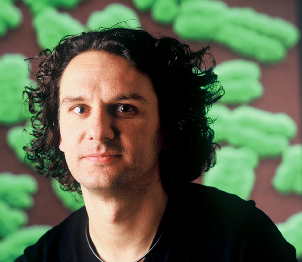
Tom Misteli ist ein international renommierter Zellbiologe, der Pionierarbeit bei der Verwendung von Bildgebungsverfahren zur Untersuchung von Genomen und Genexpression in lebenden Zellen geleistet hat. Er ist leitender Forscher und stellvertretender Direktor am National Cancer Institute (NIH). Das Interesse seines Labors besteht darin, grundlegende Prinzipien der räumlichen Genomorganisation aufzudecken und dieses Wissen für die Entwicklung neuer diagnostischer und therapeutischer Strategien für Krebs und Alterung einzusetzen. Er hat zahlreiche Auszeichnungen erhalten, darunter die Goldmedaille der Karls-Universität, den Flemming Award, den Gian-Tondury-Preis, den NIH Director's Award und einen NIH Merit Award. Er fungiert als Berater für zahlreiche nationale und internationale Agenturen und ist Mitglied mehrerer Redaktionsausschüsse, darunter Cell. Er ist Chefredakteur des Journal of Cell Biology und von Current Opinion in Cell Biology.
Das Hutchinson-Gilford-Progerie-Syndrom (HGPS) wird durch Mutationen im Lamin-A-Gen verursacht, die zur Produktion und Ansammlung eines mutierten Prälamin-A-Proteins namens Progerin führen. Da sich dieses Protein ansammelt und mit Kernkomponenten und -funktionen interferiert, ist die Identifizierung direkter Progerin-Effektoren während der Mitose und Differenzierung von entscheidender Bedeutung für das Verständnis, wie und wann Progerin die Kerndefekte auslöst, die zu vorzeitiger Alterung der Zellen führen.
In dieser Studie plant das Labor von Dr. Djabali, direkte Progerin-Effektoren innerhalb des Kerngerüsts, der Kernhülle und des Kerninneren zu identifizieren, um die anfänglichen molekularen Interaktionen zu bestimmen, die durch die Progerin-Expression gestört werden. Zu diesem Zweck werden sie Anti-Progerin-Antikörper und HGPS-Zellmodelle verwenden, darunter Fibroblasten und aus der Haut stammende Vorläuferzellen, die aus Hautbiopsien von Patienten mit HGPS (PRF Cell Bank) gewonnen wurden. Sie werden biochemische und zelluläre Bildgebung kombinieren, um Progerin-Effektoren zu identifizieren und ihren Beitrag zu den molekularen Ereignissen zu untersuchen, die zu den typischen phänotypischen Veränderungen führen, die in HGPS-Zellen beobachtet werden, die für die Entwicklung der HGPS-Krankheit verantwortlich sind. Die aus diesen Studien gewonnenen Erkenntnisse werden die Identifizierung neuer therapeutischer Ziele für die HGPS-Behandlung und neuer zellulärer Endpunkte zur Prüfung der Wirksamkeit potenzieller Eingriffe ermöglichen. Wir hoffen, dass unsere Arbeit das notwendige Wissen liefert, um uns und andere Teams im HGPS-Bereich der Suche nach einem oder mehreren Heilmitteln näher zu bringen, die Kindern mit HGPS helfen, ein längeres gesundes Leben zu führen.

Karima Djabali, PhD, ist Professorin für Epigenetik des Alterns an der Medizinischen Fakultät, Abteilung für Dermatologie und Institut für Medizintechnik (IMETUM) der Technischen Universität München. Dr. Djabali erhielt ihren MSc und PhD in Biochemie an der Universität Paris VII. Ihre Abschlussarbeit verfasste sie am College de France (Labor von Prof. F. Gros, Frankreich) und an der Rockefeller University (Labor von Prof. G. Blobel, USA). Ihre Postdoktorandenforschung führte sie am EMBL (Heidelberg, Deutschland) durch. Sie wurde 1994 zur Chargé de recherche am Nationalen Zentrum für wissenschaftliche Forschung (CNRS, Frankreich) ernannt und war von 1999 bis 2003 als assoziierte Wissenschaftlerin in der Abteilung für Dermatologie der Columbia University of New York (USA) tätig. Danach war Dr. Djabali von 2004 bis 2009 Assistenzprofessorin in der Abteilung für Dermatologie der Columbia University of New York (USA). Dr. Djabalis Forschung konzentriert sich auf die Zellalterung im Normal- und Krankheitszustand, mit besonderem Schwerpunkt auf der molekularen und zellulären Pathogenese von vorzeitigen Alterungskrankheiten wie dem Hutchinson-Gilford-Progerie-Syndrom (HGPS). Ihre Forschung kombiniert Molekularbiologie, Zellbiologie, Genetik und Proteomik, um mit der Zellalterung in Zusammenhang stehende Signalwege zu identifizieren und vorbeugende Strategien zur Verzögerung und/oder Korrektur von Alterungsprozessen zu entwickeln.
Das Labor von Dr. Misteli versucht, Leitsubstanzen für die Entwicklung von HGPS-Medikamenten durch das Screening großer Bibliotheken chemischer Moleküle zu identifizieren. Der Specialty Award wurde für den Kauf der für diese Studien erforderlichen Roboter-Laborausrüstung verwendet.
Tom Misteli ist ein international renommierter Zellbiologe, der Pionierarbeit bei der Verwendung von Bildgebungsverfahren zur Untersuchung von Genomen und Genexpression in lebenden Zellen geleistet hat. Er ist leitender Forscher und stellvertretender Direktor am National Cancer Institute, NIH. Das Interesse seines Labors besteht darin, grundlegende Prinzipien der räumlichen Genomorganisation aufzudecken und dieses Wissen für die Entwicklung neuer diagnostischer und therapeutischer Strategien für Krebs und Alterung einzusetzen. Er hat zahlreiche Auszeichnungen erhalten, darunter die Goldmedaille der Karls-Universität, den Flemming Award, den Gian-Tondury-Preis, den NIH Director's Award und einen NIH Merit Award. Er fungiert als Berater für zahlreiche nationale und internationale Agenturen und ist Mitglied mehrerer Redaktionsausschüsse, darunter Zelle. Er ist Chefredakteur von Das Journal der Zellbiologie und von Aktuelle Meinung in der Zellbiologie.
Das Hutchinson-Gilford-Progerie-Syndrom (HGPS) ist eine seltene tödliche genetische Erkrankung, die durch vorzeitiges Altern und Tod im durchschnittlichen Alter von 13 Jahren gekennzeichnet ist. Die meisten HGPS-Patienten tragen eine Mutation in LMNA Gen (das hauptsächlich Lamin A und Lamin C kodiert), das zur Produktion von „Progerin“ führt, einem abnormalen Protein, das eine toxische Farnesylmodifikation beibehält. Experimente mit Zell- und Mausmodellen von HGPS haben schlüssig gezeigt, dass die Gesamtmenge an farnesyliertem Progerin und das Verhältnis von Progerin zu reifem Lamin A den Schweregrad der Krankheit bei Progerie bestimmen und ein Schlüsselfaktor für die Lebensdauer sind. Laufende klinische Studien untersuchen daher die Wirksamkeit von Medikamenten, die die Progerin-Farnesylierung bei HGPS-Patienten hemmen. Das Hauptziel dieses Projekts ist die Entwicklung einer Methode zur routinemäßigen und genauen Quantifizierung der Progerin-Expression und ihres Farnesylierungsniveaus sowie des Verhältnisses von Progerin zu reifem Lamin A in Zellen von HGPS-Patienten. Die Messung dieser Parameter wird dazu beitragen, die Wirksamkeit von Medikamenten zu beurteilen, die auf die Progerin-Farnesylierung abzielen, sowie die Wirksamkeit zukünftiger Strategien zur Hemmung der abnormalen Verarbeitung (Spleißen) des LMNA mRNA, die Ursache von HGPS bei den meisten Patienten. Ein sekundäres Ziel ist die Durchführung von Pilotstudien zur Entwicklung einer Hochdurchsatzstrategie zur Identifizierung von Mechanismen, die abweichende LMNA Spleißen.
Vicente Andrés promovierte 1990 in Biowissenschaften an der Universität Barcelona. Während seiner Postdoc-Ausbildung am Kinderkrankenhaus der Harvard University (1991-1994) und am St. Elizabeth's Medical Center der Tufts University (1994-1995) leitete er Studien zur Rolle von Homöobox- und MEF2-Transkriptionsfaktoren in Prozessen der Zelldifferenzierung und -vermehrung. In dieser Zeit entwickelte er auch sein Interesse an der Herz-Kreislauf-Forschung. Seine Karriere als unabhängiger Wissenschaftler begann 1995, als er zum Assistenzprofessor für Medizin an der Tufts University ernannt wurde. Seitdem haben Dr. Andrés und seine Gruppe die Gefäßumgestaltung bei Arteriosklerose und Restenose nach Angioplastie untersucht. In jüngster Zeit untersuchen sie die Rolle der Kernhülle bei der Regulierung der Signalübertragung, Genexpression und Zellzyklusaktivität bei Herz-Kreislauf-Erkrankungen und Alterung, mit besonderem Schwerpunkt auf A-Typ-Laminen und dem Hutchinson-Gilford-Progerie-Syndrom (HGPS).
Nachdem er eine Stelle als fest angestellter Wissenschaftler am spanischen Nationalen Forschungsrat (CSIC) erhalten hatte, kehrte Dr. Andrés 1999 nach Spanien zurück, um seine Forschungsgruppe am Institut für Biomedizin in Valencia aufzubauen, wo er als ordentlicher Professor arbeitete. Seit 2006 ist seine Gruppe Mitglied der Red Temática de Investigación Cooperativa en Enfermedades Cardiovasculares (RECAVA). Im September 2009 trat er dem Centro Nacional de Investigaciones Cardiovasculares (CNIC) bei. 2010 wurde ihm von der belgischen Gesellschaft für Kardiologie der Doktor Leon Dumont-Preis verliehen.
 Dr. Benchimol hat eine lange Erfolgsgeschichte auf dem Gebiet der p53-Funktion vorzuweisen. Er wird sein Fachwissen nutzen, um auf faszinierenden vorläufigen Daten aufzubauen und neue Hypothesen über die Rolle von p53 bei der Vermittlung der vorzeitigen Seneszenz zu testen, die Zellen von Patienten mit Hutchinson-Gilford-Progerie-Syndrom (HGPS) aufweisen. Das erste Ziel besteht darin, die Hypothese zu testen, dass Progerin Replikationsstress verursacht, der wiederum einen Wachstumsstopp der Seneszenz auslöst, und dass p53 nach dem durch Progerin verursachten Replikationsstress wirkt. Auf dieses Ziel folgt ein mechanistischeres Ziel, das bestimmen soll, wie Progerin und p53 zusammenarbeiten, um eine Seneszenzreaktion hervorzurufen.
Juli 2012: An Tom Misteli, PhD, National Cancer Institute, NIH, Bethesda, MD; Änderung des Specialty Award
Das Labor von Dr. Misteli versucht, Leitsubstanzen für die Entwicklung von HGPS-Medikamenten durch das Screening großer Bibliotheken chemischer Moleküle zu identifizieren. Der Specialty Award wurde für den Kauf der für diese Studien erforderlichen Roboter-Laborausrüstung verwendet.
Tom Misteli ist ein international renommierter Zellbiologe, der Pionierarbeit bei der Verwendung von Bildgebungsverfahren zur Untersuchung von Genomen und Genexpression in lebenden Zellen geleistet hat. Er ist leitender Forscher und stellvertretender Direktor am National Cancer Institute, NIH. Das Interesse seines Labors besteht darin, grundlegende Prinzipien der räumlichen Genomorganisation aufzudecken und dieses Wissen für die Entwicklung neuer diagnostischer und therapeutischer Strategien für Krebs und Alterung einzusetzen. Er hat zahlreiche Auszeichnungen erhalten, darunter die Goldmedaille der Karls-Universität, den Flemming Award, den Gian-Tondury-Preis, den NIH Director's Award und einen NIH Merit Award. Er fungiert als Berater für zahlreiche nationale und internationale Agenturen und ist Mitglied mehrerer Redaktionsausschüsse, darunter Zelle. Er ist Chefredakteur von Das Journal der Zellbiologie und von Aktuelle Meinung in der Zellbiologie.
 A-Typ-Laminine sind wichtige Strukturproteine des Zellkerns von Säugetierzellen. Sie sind die Hauptbestandteile eines filamentartigen Geflechts an der Innenseite der Kernhülle und verleihen dem Zellkern nicht nur Form und mechanische Stabilität, sondern sind auch an wesentlichen zellulären Prozessen wie der DNA-Replikation und Genexpression beteiligt. Neben ihrer Lokalisierung an der Kernperipherie ist im Inneren des Kerns ein zusätzlicher, dynamischerer Pool von A-Typ-Laminen vorhanden, der vermutlich für die ordnungsgemäße Zellvermehrung und -differenzierung wichtig ist. In den letzten dreizehn Jahren wurden über 300 Mutationen im Gen, das A-Typ-Laminine kodiert, mit verschiedenen menschlichen Krankheiten in Verbindung gebracht, darunter auch mit der vorzeitigen Alterungskrankheit Hutchinson-Gilford-Progerie-Syndrom (HGPS). Die molekularen Krankheitsmechanismen sind noch immer schlecht verstanden, was die Entwicklung wirksamer therapeutischer Strategien behindert. Die mit HGPS verbundene Mutation im A-Typ-Lamin-Gen führt zur Produktion eines mutierten Lamin-A-Proteins, das als Progerin bezeichnet wird. Im Gegensatz zu normalem Lamin A ist Progerin stabil in der Kernmembran verankert, was die mechanischen Eigenschaften des Kerns verändert. Unsere Arbeitshypothese geht davon aus, dass das membranverankerte Progerin auch den dynamischen Pool an Laminen im Inneren des Kerns und damit die Zellproliferation und -differenzierung stark beeinflusst.
Ein Ziel dieses Projekts ist es, die Mechanismen zu identifizieren, die für die Verankerung von Progerin an der Kernmembran verantwortlich sind, und Wege zu finden, diese Membranverankerung gezielt zu hemmen, mit der Aussicht, den dynamischen Laminpool zu retten und dadurch die mit HGPS verbundenen zellulären Phänotypen umzukehren. Frühere Erkenntnisse zeigen, dass dieser dynamische Laminpool in einem Komplex mit anderen Proteinen die Zellproliferation über den Retinoblastomprotein-(pRb)-Signalweg reguliert. Zur Unterstützung unserer Hypothese wurde kürzlich gezeigt, dass der pRb-Signalweg in Zellen von HGPS-Patienten tatsächlich beeinträchtigt ist. Im zweiten Ziel unseres Projekts schlagen wir vor, die Auswirkungen von Progerin auf die Regulierung, Dynamik und Aktivitäten des mobilen, nukleoplasmatischen Lamin-A-Pools und seiner assoziierten Proteine sowie seine Auswirkungen auf die pRb-Signalgebung im molekularen Detail zu untersuchen. Die Ergebnisse unserer Studie werden voraussichtlich Licht auf die krankheitsverursachenden molekularen Mechanismen hinter HGPS werfen und möglicherweise dazu beitragen, neue Wirkstoffziele und Medikamente für effizientere und gezieltere Therapien zu identifizieren.
Dr. Dechat erhielt seinen MSc und PhD in Biochemie an der Universität Wien, Österreich. Nach einem Jahr als PostDoc an der Abteilung für neuromuskuläre Forschung der Medizinischen Universität Wien war er von 2004 bis 2009 PostDoc im Labor von Prof. Robert Goldman, Northwestern University, Feinberg Medical School, Chicago, Illinois, wo er an der strukturellen und funktionellen Charakterisierung von Kernlaminen im gesunden und kranken Zustand arbeitete, mit einem Schwerpunkt auf den Mechanismen, die aufgrund der Expression von Progerin zum Hutchinson-Gilford-Progerie-Syndrom führen. Seit 2010 ist er Assistenzprofessor an den Max F. Perutz Laboratories der Medizinischen Universität Wien und untersucht die strukturellen und funktionellen Eigenschaften von nukleoplasmatischen A-Typ-Lamina und LAP2 während des Zellzyklus und bei verschiedenen Krankheiten, die mit Mutationen in den Laminen A/C und LAP2 in Zusammenhang stehen.
 In dieser Studie plant Dr. Erikssons Labor, sein kürzlich entwickeltes Modell für Progerie mit Expression der häufigsten LMNA-Genmutation im Knochen zu verwenden. Sie haben zuvor gezeigt, dass die Unterdrückung der Expression der Progerie-Mutation nach der Entwicklung der Progerie-Hautkrankheit zu einer fast vollständigen Umkehrung des Krankheitsphänotyps führte (Sagelius, Rosengardtenet al. 2008). Der Krankheitsverlauf der Progerie wird zu verschiedenen Zeitpunkten nach der Hemmung der Mutation im Knochengewebe überwacht, um die Möglichkeit einer Umkehrung der Krankheit zu analysieren. Ihre vorläufigen Ergebnisse deuten auf eine Verbesserung der klinischen Symptome hin und geben Anlass zur Hoffnung, eine mögliche Behandlung und Heilung für diese Krankheit zu finden.
Dr. Eriksson erhielt 1996 ihren MSc in Molekularbiologie an der Universität Umeå, Schweden, und 2001 ihren PhD in Neurologie vom Karolinska Institutet. Von 2001 bis 2003 war sie Postdoktorandin am National Human Genome Research Institute, National Institutes of Health, und ist seit 2003 PI/Forschungsgruppenleiterin und Assistenzprofessorin an der Abteilung für Biowissenschaften und Ernährung am Karolinska-Institut. Sie ist außerdem außerordentliche Professorin für medizinische Genetik am Karolinska-Institut. Ihre Forschungsinteressen umfassen Progerie und genetische Mechanismen des Alterns.
Dezember 2011 (Startdatum 1. März 2012): An Colin L. Stewart D.Phil, Institute of Medical Biology, Singapur; „Definition der molekularen Grundlagen der Verschlechterung der Gefäßglattmuskulatur bei Progerie
Kinder mit Progerie sterben an Herz-Kreislauf-Erkrankungen, entweder einem Herzinfarkt oder einem Schlaganfall. Im letzten Jahrzehnt wurde deutlich, dass die Blutgefäße des Kindes ein wichtiges von Progerie betroffenes Gewebe sind. Progerie scheint die Muskelwand der Blutgefäße zu schwächen, indem sie auf irgendeine Weise das Absterben der glatten Muskelzellen verursacht. Dies kann die Gefäße nicht nur fragiler machen, sondern auch die Bildung von Plaque fördern, was zu einer Verstopfung des Gefäßes führt. Beide Folgen führen zum Versagen der Blutgefäße und, wenn dies die Herzgefäße betrifft, führt dies zu einem Herzinfarkt.
Colin Stewart und sein Kollege Oliver Dreesen wollen untersuchen, wie die defekte Form des Kernproteins Lamin A (Progerin) speziell das Wachstum und Überleben der glatten Muskelzellen in Blutgefäßen beeinflusst. Mithilfe der Stammzellentechnologie konnten Colin und seine Kollegen Stammzellen aus Hautzellen von zwei Kindern mit Progerie gewinnen. Diese patientenspezifischen Stammzellen wurden dann in glatte Muskelzellen umgewandelt, die denen aus Blutgefäßen ähneln. Interessanterweise produzierten diese glatten Muskelzellen im Vergleich zu anderen Zelltypen einige der höchsten Progerinwerte, was einen möglichen Grund dafür nahelegt, warum Blutgefäße bei Progerie so stark betroffen sind. Glatte Muskelzellen mit Progerin zeigten Anzeichen einer Schädigung der DNA im Zellkern. Colin und Oliver werden diese und andere aus den Stammzellen gewonnene Zellen verwenden, um zu verstehen, welche Art von DNA beschädigt ist und welche biochemischen Prozesse, die für das Überleben der glatten Muskelzellen notwendig sind, durch Progerin beeinflusst werden. Durch die direkte Untersuchung von glatten Muskelzellen von Kindern mit Progerie hoffen sie, genau herauszufinden, was mit den Zellen nicht stimmt. Auf diese Weise können sie neuartige Verfahren zum Testen neuer Medikamente entwickeln, die möglicherweise irgendwann bei der Behandlung betroffener Kinder helfen könnten.
Colin Stewart erhielt seinen Doktortitel von der Universität Oxford, wo er die Wechselwirkungen zwischen Teratokarzinomen, den Vorläufern von ES-Zellen, und frühen Mausembryonen untersuchte. Nach seiner Postdoc-Arbeit bei Rudolf Jaenisch in Hamburg war er wissenschaftlicher Mitarbeiter am EMBL in Heidelberg. Dort war er maßgeblich an der Entdeckung der Rolle des Zytokins LIF bei der Erhaltung von Maus-ES-Zellen beteiligt. Außerdem begann er sich für die Kernlaminen und die Kernarchitektur während der Entwicklung zu interessieren. Nach seinem Wechsel an das Roche Institute of Molecular Biology in New Jersey setzte er seine Studien zu den Laminen, Stammzellen und genomischer Prägung fort. 1996 wechselte er zum ABL-Forschungsprogramm in Frederick, Maryland, und wurde 1999 zum Leiter des Labors für Krebs- und Entwicklungsbiologie am National Cancer Institute ernannt. Im letzten Jahrzehnt konzentrierten sich seine Interessen auf die funktionelle Architektur des Zellkerns in Stammzellen, Regeneration, Alterung und Krankheit, insbesondere im Hinblick darauf, wie die Kernfunktionen in die Dynamik des Zytoskeletts während der Entwicklung und bei Krankheiten integriert sind. Seit Juni 2007 ist er leitender Forschungsleiter und stellvertretender Direktor am Institute of MedicalBiology der Singapore Biopolis.
Oliver Dreesen ist derzeit Senior Research Fellow am Institute of Medical Biology in Singapur. Nach Abschluss seines Bachelor-Studiums in Bern, Schweiz, war Oliver als Forscher am Pasteur Institute in Paris und an der University of California in San Diego tätig. Er promovierte an der Rockefeller University in New York, wo er die Struktur und Funktion von Chromosomenenden (Telomeren) während der antigenen Variation bei afrikanischen Trypanosomen untersuchte. Sein aktueller Forschungsschwerpunkt liegt auf der Rolle der Telomere bei menschlichen Krankheiten, Alterung und zellulärer Neuprogrammierung.
 Das Hutchinson-Gilford-Progerie-Syndrom (HGPS) ist eine seltene und schwächende Krankheit, die durch eine Mutation im Lamin-A-Protein verursacht wird. Frühere Studien haben die Mutationen in Lamin A identifiziert, die die Krankheit verursachen, und ihre abweichende Funktion in menschlichen Zellen und in Mausmodellen von HGPS untersucht. Diese Informationen, zusammen mit genomweiten Expressionsstudien, bei denen HGPS-Zellen mit denen von nicht betroffenen Personen verglichen wurden, haben unser Verständnis dieser Krankheit dramatisch erweitert. Ein Bereich, der in der HGPS-Forschung vernachlässigt wurde, ist eine gründliche Analyse der Stoffwechselveränderungen, die in HGPS-Zellen im Vergleich zu gesunden Kontrollpersonen auftreten. Stoffwechselstörungen begleiten viele menschliche Krankheiten (z. B. Arteriosklerose, Diabetes und Krebs), und die klinische Bewertung von HGPS deutet auf chronische Anomalien in grundlegenden Stoffwechselwegen hin.
Zelluläre Metabolite stellen die Biochemikalien dar, die – zusammen mit Proteinen und Nukleinsäuren – das gesamte Repertoire an Molekülen in einer Zelle bilden. Daher sind Stoffwechselveränderungen bei der Krankheitsentstehung wohl ebenso wichtig wie Veränderungen der Genexpression. Tatsächlich hat das aufstrebende Feld der „Metabolomik“ bereits viele wichtige Entdeckungen hervorgebracht, die Folgendes verbinden: einzelne Metabolite auf bestimmte menschliche Krankheiten, darunter Leukämie und metastasierenden Prostatakrebs. Daher sollte die Identifizierung der Metaboliten und Stoffwechselwege, die bei HGPS verändert sind, Einblicke in die Krankheitspathogenese geben und möglicherweise völlig neue therapeutische Strategien aufdecken. Dies ist insbesondere für HGPS von Bedeutung, da zahlreiche zellbasierte und In-vivo-Studien gezeigt haben, dass Lamin-A-Mutationen keine irreversiblen Schäden verursachen und dass zelluläre HGPS-Phänotypen bei richtiger Behandlung tatsächlich eliminiert werden können.
Nach Abschluss eines umfassenden, vergleichenden Screenings der in Zellen von gesunden Spendern und HGPS-Patienten vorhandenen Metaboliten werden nachfolgende biochemische und zellbasierte Tests feststellen, ob die im Screening identifizierten Schlüsselmetaboliten HGPS-Phänotypen in gesunden Zellen auslösen oder HGPS-Phänotypen in erkrankten Zellen umkehren können. Folglich wird diese Studie nicht nur aufdecken, wie HGPS-assoziierte Lamin-A-Mutationen globale Stoffwechselwege in menschlichen Zellen beeinflussen, sondern auch beginnen zu bewerten, ob das gezielte Angreifen dieser Wege einen wirksamen Ansatz für therapeutische Eingriffe darstellt.
Das Taatjes-Labor kombiniert Fachwissen in Biochemie, Proteomik und Kryo-Elektronenmikroskopie, um die grundlegenden Mechanismen zu untersuchen, die die Genexpression des Menschen regulieren. Das Labor implementiert auch genomweite und metabolomische Ansätze, um mechanistische Erkenntnisse mit physiologischen Konsequenzen zu verknüpfen. Metabolomische Studien im Taatjes-Labor dienen in Verbindung mit mechanistischen Studien mit einer p53-Isoform, die eine beschleunigte Alterung verursacht, als Grundlage für diese HGPS-Studie.
 Das Hutchinson-Gilford-Progerie-Syndrom (HGPS) wird durch Mutationen im Gen verursacht, das die Laminine A und C kodiert. Kinder mit HGPS entwickeln Haarausfall, Knochendefekte, Verlust von Fettgewebe und andere Zeichen beschleunigter Alterung, bevor sie im frühen Teenageralter einen Schlaganfall oder Herzinfarkt erleiden. Postmortem-Studien zeigen einen dramatischen Verlust von vaskulären glatten Muskelzellen in den größeren Blutgefäßen von HGPS-Patienten. Gefäßglatte Muskelzellen sind für die normale Funktion der Blutgefäße von entscheidender Bedeutung, und der Verlust von Gefäßglatte Muskelzellen könnte die treibende Kraft hinter der tödlichen Herz-Kreislauf-Erkrankung bei HGPS sein.
Wir haben bereits gezeigt, dass Hautzellen von HGPS-Patienten empfindlicher auf mechanische Belastungen reagieren, was zu einem erhöhten Zelltod bei wiederholter Dehnung führt. In diesem Projekt werden wir untersuchen, ob eine erhöhte Empfindlichkeit gegenüber mechanischer Belastung auch für den fortschreitenden Verlust von vaskulären glatten Muskelzellen bei HGPS verantwortlich ist, da große Blutgefäße bei jedem Herzschlag wiederholter Gefäßbelastung ausgesetzt sind. In Kombination mit einer beeinträchtigten Erneuerung der beschädigten Zellen könnte die erhöhte mechanische Empfindlichkeit zum fortschreitenden Verlust von vaskulären glatten Muskelzellen und zur Entwicklung von Herz-Kreislauf-Erkrankungen bei HGPS führen.
Um die Wirkung von mechanischem Stress auf vaskuläre glatte Muskelzellen in vivo zu untersuchen, werden wir chirurgische Verfahren anwenden, um den Blutdruck lokal zu erhöhen oder Gefäßverletzungen in großen Blutgefäßen zu verursachen. Anschließend vergleichen wir die Wirkung auf das Überleben und die Regeneration von vaskulären glatten Muskelzellen in einem Mausmodell von HGPS und in gesunden Kontrollpersonen. Die Erkenntnisse aus diesen Studien werden neue Informationen über die molekularen Mechanismen liefern, die der Herz-Kreislauf-Erkrankung bei HGPS zugrunde liegen, und könnten neue Hinweise für die Entwicklung therapeutischer Ansätze liefern.
Dr. Lammerding ist Assistenzprofessor an der Cornell University im Fachbereich Biomedizintechnik und am Weill Institute for Cell and Molecular Biology. Bevor er 2011 an die Cornell University wechselte, arbeitete Dr. Lammerding als Assistenzprofessor im Fachbereich Medizin der Harvard Medical School/Brigham and Women's Hospital und war Dozent am Massachusetts Institute of Technology. Das Lammerding-Labor untersucht die subzelluläre Biomechanik und die zelluläre Signalreaktion auf mechanische Stimulation, mit besonderem Schwerpunkt darauf, wie Mutationen in Kernhüllproteinen wie Laminen Zellen empfindlicher gegenüber mechanischem Stress machen und ihre Mechanotransduktionssignalisierung beeinflussen können. Die aus dieser Arbeit gewonnenen Erkenntnisse können zu einem besseren Verständnis der molekularen Mechanismen führen, die verschiedenen Laminopathien zugrunde liegen, einer vielfältigen Gruppe von Krankheiten, zu denen das Hutchison-Gilford-Progerie-Syndrom, die Emery-Dreifuss-Muskeldystrophie und die familiäre partielle Lipodystrophie gehören.
Die Kernlaminate vom Typ A und B sind Proteine, die sich im Zellkern befinden. Diese Proteine bilden getrennte, aber interagierende Strukturnetzwerke im Zellkern. Die Laminate sind für die Bestimmung der Größe, Form und mechanischen Eigenschaften des Zellkerns von entscheidender Bedeutung und sie bilden ein intranukleäres Gerüst für die Organisation der Chromosomen. Wir haben entdeckt, dass, wenn ein Laminate-Netzwerk durch eine Mutation verändert wird, die zu einer Fehlfunktion führt, auch das andere verändert wird. Obwohl die typischen und atypischen Formen des Hutchinson-Gilford-Progerie-Syndroms durch unterschiedliche Mutationen im Kernlaminat-A-Gen verursacht werden, haben wir festgestellt, dass die Laminate-Netzwerke vom Typ B in den Zellen von Progeriepatienten ebenfalls abnormal verändert sind. Die Laminate vom Typ B werden ab der Befruchtung in allen somatischen Zellen exprimiert und sind bekanntermaßen wichtig für die Regulierung vieler Kernfunktionen, einschließlich der DNA-Replikation und der Gentranskription. Den Laminate-B-Isoformen und ihrer Rolle bei der Progerie wurde jedoch wenig Aufmerksamkeit geschenkt. In diesem Vorschlag ist es unser Ziel, die Auswirkungen der Expression von Progerin, der am häufigsten vorkommenden mutierten Form von Lamin A, und anderer atypischer Progerie-Lamin A-Mutationen auf die Expression, Struktur und Funktion der B-Typ-Laminine zu bestimmen. Unsere vorläufigen Studien legen nahe, dass Veränderungen in den B-Typ-Lamin-Netzwerken aufgrund ihrer Wechselwirkungen mit A-Typ-Lamininen wichtige Mediatoren der Zellpathologie bei HGPS sind. Wir werden Veränderungen in den B-Typ-Lamininen in Zellen von Progeriepatienten und ihre Beziehung zu Zellwachstumsdefekten und vorzeitiger Seneszenz untersuchen. Wir werden auch die Auswirkungen der Hemmung der Farnesyltransferase auf die Expression, Modifikation und Stabilität der B-Typ-Laminine untersuchen. Dies ist wichtig, da B-Typ-Laminine normalerweise stabil farnesyliert sind. Diese vorgeschlagenen Studien kommen angesichts der laufenden klinischen Studien mit Progeriepatienten, die Medikamente verwenden, die die Proteinfarnesylierung hemmen, besonders zur rechten Zeit. Unsere Studien versprechen neue Einblicke in die molekularen Mechanismen, die für die vorzeitige Alterung der Zellen bei Patienten mit dieser verheerenden Krankheit verantwortlich sind. Die Ergebnisse unserer Untersuchungen sollten Erkenntnisse über zusätzliche potenzielle Angriffspunkte liefern, die bei der Entwicklung neuer Therapien für HGPS-Patienten berücksichtigt werden müssen.
Robert D. Goldman, PhD, ist Stephen Walter Ranson Professor und Vorsitzender der Abteilung für Zell- und Molekularbiologie an der Feinberg School of Medicine der Northwestern University in Chicago. Dr. Goldman erhielt seinen Doktortitel in Biologie von der Princeton University und führte Postdoc-Forschungen an der University of London und am MRC Institute of Virology in Glasgow durch. Er war an der Case Western Reserve University und der Carnegie-Mellon University tätig und Gastwissenschaftler am Cold Spring Harbor Laboratory, bevor er zu Northwestern kam. Er gilt als anerkannte Autorität auf dem Gebiet der Struktur und Funktion der nukleoskelettalen und zytoskelettalen Intermediärfilamentsysteme. Anfang der 1980er Jahre war er fasziniert von der Entdeckung, dass Laminen die Kernform der Intermediärfilamente sind. Seitdem hat sein Forschungslabor gezeigt, dass die Kernlaminen die Größe und Form des Zellkerns bestimmen und dass sie bei der Zerlegung und Rekonstruktion des Zellkerns während der Zellteilung von entscheidender Bedeutung sind. Seine Forschungsgruppe hat außerdem gezeigt, dass sich die Lamine im Zellkern zu einem molekularen Gerüst zusammensetzen, das für die DNA-Replikation, Transkription und Chromatinorganisation erforderlich ist. In den letzten Jahren konzentrierte sich sein Interesse an den Laminen auf die Auswirkungen von Lamin-A-Mutationen, die zu der vorzeitigen Alterungskrankheit Hutchinson-Gilford-Progerie-Syndrom und anderen atypischen Formen der Progerie führen. Dies führte seine Forschungen zur Bestimmung der Rolle der Lamine bei der Chromosomenorganisation, bei der Regulierung der epigenetischen Modifikationen des Chromatins sowie bei der Zellproliferation und -alterung.
Dr. Goldman ist Fellow der American Association for the Advancement of Science (AAAS) und wurde mit dem Ellison Medical Foundation Senior Scholar Award und dem NIH MERIT Award ausgezeichnet. Er ist ein produktiver Autor, hat zahlreiche Bände für den Cold Spring Harbor Laboratory Press herausgegeben und ist Mitherausgeber des FASEB Journal und von Molecular Biology of the Cell. Er wurde in zahlreiche wissenschaftliche Gesellschaften gewählt, darunter in den Vorstand der AAAS, den Rat und Präsidenten der American Society for Cell Biology, und war Präsident der American Association of Anatomy, Cell Biology and Neuroscience Chairs. Er war Mitglied zahlreicher Prüfungsausschüsse der American Cancer Society und der NIH, ist Direktor des Whitman Center of the Marine Biological Laboratory und wird häufig eingeladen, internationale Tagungen im In- und Ausland zu organisieren und dort Vorträge zu halten.
Das Hutchinson-Gilford-Progerie-Syndrom (HGPS) wird durch eine Mutation im Lamin-A-Gen verursacht, die zur Produktion und Ansammlung des mutierten Krankheitsproteins Progerin führt. Da sich dieses Protein ansammelt, ist es aus therapeutischer Sicht wichtig, herauszufinden, wie es abgebaut wird. Der Schwerpunkt dieser Arbeit liegt auf der Bestimmung der zellulären Abbauwege, die für den Abbau des Progerin-Proteins verantwortlich sind. Mithilfe dieser Informationen hoffen wir, diese Wege manipulieren zu können, um den Abbau von Progerin zu erleichtern, mit dem Ziel, aktuelle oder zukünftige Therapien für HGPS zu verbessern.
Dr. Graziotto ist Postdoktorand in der Abteilung für Neurologie am Massachusetts General Hospital. Derzeit arbeitet er im Labor von Dr. Dimitri Krainc. Ein Schwerpunkt des Labors ist die Erforschung neurodegenerativer Erkrankungen, bei denen sich mutierte Proteine ansammeln und Aggregate bilden. Das Labor untersucht die Clearance-Mechanismen dieser Proteine, um Modifikatoren dieser Signalwege zu identifizieren, die zu zukünftigen Behandlungszielen führen könnten.
 „Progerie“ beschreibt eine Reihe von Erkrankungen, die unterschiedliche Aspekte vorzeitiger Alterung oder segmentaler Progerie aufweisen. Dazu gehören HGPS und MAD, beide mit LMNA-Mutationen, sowie die DNA-Reparaturstörungen Cockayne- und Werner-Syndrom. Darüber hinaus gibt es eine Reihe von Fällen „atypischer“ Progerie mit überlappenden, aber unterschiedlichen Merkmalen. PRF hat Zelllinien und/oder DNA von 12 solcher Fälle atypischer Progerie gesammelt, was die größte jemals zusammengestellte Kohorte darstellt. DNAs wurden auf LMNA-Exon-Mutationen untersucht und es wurden keine gefunden. Derzeit werden sie in Dr. Glovers Labor auf ZMPSTE-Mutationen getestet. Darüber hinaus weisen sie Phänotypen auf, die sich von den klassischen Werner- und Cockayne-Syndromen unterscheiden. Daher haben diese Personen Mutationen in einzigartigen Progerie-Genen. Da die meisten dieser Fälle sporadisch sind, war dies eine gewaltige Aufgabe. In den letzten Jahren wurden jedoch enorme technische Fortschritte im Bereich der DNA-Sequenzierung erzielt. Die Exonsequenzierung des gesamten Genoms oder „Exomsequenzierung“ wurde erfolgreich eingesetzt, um mutierte Gene für eine Reihe monogener Merkmale zu identifizieren, darunter das Miller-Syndrom, das Kabuki-Syndrom, die unspezifische geistige Behinderung, das Perrault-Syndrom und viele andere. Zahlreiche weitere Studien sind im Gange, darunter viele Studien über neu Mutationen. Dies ist ein leistungsfähiges Werkzeug zur Genidentifizierung und es wird vorausgesagt, dass wir in den nächsten Jahren die genetische Ursache der meisten monogenen Merkmale verstehen werden.
Angesichts dieser technologischen Fortschritte und der Verfügbarkeit ähnlicher Patienten vermutet Dr. Glover, dass Mutationen, die für atypische Progerie verantwortlich sind, durch eine vollständige Exomsequenzierung dieser Patientenproben identifiziert werden können. Die Identifizierung dieser Mutationen ist für das Verständnis der Krankheitsätiologie, die Entwicklung wirksamer Behandlungen und für die Entwicklung von Kenntnissen über sich kreuzende und interagierende molekulare und zelluläre Wege bei Progerie und normalem Altern von entscheidender Bedeutung. Dies ist jedoch eine Herausforderung, da es sich anscheinend alles um Neumutationen handelt und die Phänotypen heterogen sind. Das unmittelbare Ergebnis dieser Studie wird die Entdeckung von 7-15 neuen, wahrscheinlich schädlichen Mutationen für jede Familie sein, die von betroffenen Familienmitgliedern geteilt werden und für die Familie einzigartig sein können. Die gemeinsame Analyse dieser Gene über 6-12 Familien hinweg kann durchaus Fälle unterschiedlicher schädlicher Allele desselben Gens oder verschiedener Defekte im selben Funktionsweg aufdecken, die in mehreren Familien auftreten, und so den ersten Einblick in neue Kandidatengene/-wege für Progerie geben. Im Erfolgsfall könnten die Erkenntnisse enorme Auswirkungen haben und nicht nur für den betroffenen Patienten, sondern aufgrund überlappender Merkmale auch für andere Formen der Progerie, einschließlich HGPS, sowie für die normale Alterung von direkter Bedeutung sein.
Dr. Glover ist Professor in der Abteilung für Humangenetik und Pädiatrie an der University of Michigan. Er ist Autor von über 120 Forschungspublikationen und Buchkapiteln. Dr. Glover ist seit über einem Jahrzehnt aktiv in der Progerieforschung tätig und ist seit seiner Gründung im Jahr 2004 Mitglied des PRF Medical Research Committee. Sein Labor war an den Forschungsbemühungen beteiligt, bei denen erstmals LMNA-Genmutationen bei HGPS identifiziert wurden, und an der Demonstration, dass Farnysylierungsinhibitoren die Kernanomalien von HGPS-Zellen rückgängig machen können, was die Tür für klinische Studien öffnet. Ein Hauptinteresse seines Labors gilt den Mechanismen und Folgen der Genominstabilität bei genetischen Erkrankungen des Menschen. Die aktuellen Bemühungen zielen darauf ab, die molekularen Mechanismen zu verstehen, die bei der Entstehung von Mutationen der Kopienzahlvarianten (CNV) im menschlichen Genom eine Rolle spielen. Dies ist eine häufige, aber erst kürzlich erkannte Mutationsform, die bei normalen menschlichen Variationen und zahlreichen genetischen Erkrankungen wichtig ist. Im Gegensatz zu anderen Mutationsformen ist jedoch nicht vollständig verstanden, wie sie gebildet werden und welche genetischen und umweltbedingten Risikofaktoren damit verbunden sind.
Das Ziel dieses Projekts ist es, die molekulare Grundlage der Replikationsanomalie und Genominstabilität in Zellen des Hutchinson-Gilford-Progerie-Syndroms (HGPS) zu definieren. HGPS ist eine vorherrschende Erkrankung vorzeitiger Alterung, und die durchschnittliche Lebenserwartung der Patienten beträgt nur 13 Jahre. Die Krankheit wird durch eine Punktmutation an Position 1822 oder 1824 im Exon 11 des Lamin-A-Gens verursacht, die zur sporadischen Produktion eines mutierten Lamin-A-Proteins mit 50 intern verkürzten Aminosäuren namens Progerin führt. Lamin A ist ein wichtiger innerer Bestandteil der Kernhülle und des Kernskeletts von Zellen, und die Anwesenheit von Progerin führt zu einer abnormalen Kernmorphologie und Genominstabilität in HGPS-Zellen. Interessanterweise haben neuere Studien gezeigt, dass Progerin auch bei normal alternden Personen produziert wird und sein Spiegel in den Koronararterien mit zunehmendem Alter um durchschnittlich 3% pro Jahr anzusteigen scheint. Dieser Anstieg steht im Einklang mit vielen Aspekten der Herz-Kreislauf-Pathologie sowohl bei HGPS- als auch bei geriatrischen Patienten und weist auf eine potenziell wichtige Rolle von Progerin bei der Alterung und altersbedingten Krankheiten wie Krebs und Herz-Kreislauf-Erkrankungen hin.
Während die genetische Ursache von HGPS bekannt ist, sind die molekularen Mechanismen, durch die die Wirkung von Progerin zu vorzeitigen Alterungsphänomenen führt, noch lange nicht klar. Wir und andere haben kürzlich gezeigt, dass HGPS einen Phänotyp der Genominstabilität aufweist, der durch die zelluläre Ansammlung von DNA-Doppelstrangbrüchen (DSBs) verursacht wird. Die Ansammlung von DSBs ist auch eine häufige Ursache für systemisches Altern. Wir haben auch festgestellt, dass Xeroderma pigmentosum Gruppe A (XPA) lokalisiert sich fehllokalisiert an DSB-Stellen in HGPS-Zellen, was zu einer Hemmung der DSB-Reparatur führt. Der Abbau von XPA in HGPS-Zellen stellt die DSB-Reparatur teilweise wieder her. Basierend auf diesen Erkenntnissen vermuten wir, dass die Ansammlung von DNA-Schäden in HGPS wahrscheinlich auf abweichende Aktivitäten an Replikationsgabeln zurückzuführen ist, die irreparable DSBs erzeugen, was zu einem frühen Replikationsstopp oder einer replikativen Seneszenz führt. Angesichts der Tatsache, dass HPGS-Zellen durch einen frühen Replikationsstopp und eine vorzeitige replikative Seneszenz gekennzeichnet sind, könnte die Aufklärung der Mechanismen, die den fehlerhaften Aktivitäten an Replikationsgabeln zugrunde liegen, ein Schlüssel zum Verständnis der Ursachen von HGPS-Phänotypen sein. Das Verständnis könnte zu neuen Strategien zur Behandlung der Krankheit führen, indem in die krankheitsverursachenden molekularen Wege eingegriffen wird. Andererseits ist bekannt, dass HGPS-Patienten anscheinend krebsfrei sind. Obwohl der Mechanismus unbekannt bleibt, kann er auf die vorzeitige replikative Seneszenz von HPGS zurückgeführt werden. In diesem Forschungsprojekt werden wir die molekulare Basis der DSB-Akkumulation in HGPS bestimmen und dabei den Schwerpunkt auf das Verständnis legen, wie DNA-Schäden an Replikationsgabeln entstehen. Als nächstes werden wir feststellen, ob Progerin mit DNA-Replikationsfaktoren interagiert und wie diese Interaktion Replikationsanomalien verursacht.
Dr. Zou ist Professor in der Abteilung für Biochemie und Molekularbiologie des Quillen College of Medicine an der East Tennessee State University. Er erhielt 1991 seinen Doktortitel in Biophysik von der Clark University. Dr. Zous Forschung konzentriert sich hauptsächlich auf das Verständnis der Genominstabilität bei Krebs und verwandter Prozesse, einschließlich DNA-Reparatur und DNA-Schadenskontrollpunkten. Er interessiert sich seit kurzem für Genominstabilität und DNA-Schadensreaktionen bei Progerie, die durch fehlerhafte Reifung von Prelamin A verursacht werden, insbesondere das Hutchinson-Gilford-Progerie-Syndrom, und seine Gruppe hat interessante Erkenntnisse über die molekularen Mechanismen der Genominstabilität bei HGPS gewonnen.
Dr. Cao wird in seiner Arbeit die Wirkung von Everolimus auf HGPS-Zellen untersuchen, allein oder in Kombination mit Lanafarnib. Diese Studie wird die Bewertung sowohl des therapeutischen Potenzials als auch der mechanistischen Grundlagen für einen solchen kombinatorischen therapeutischen Ansatz ermöglichen.
Dr. Cao ist Assistenzprofessor in der Abteilung für Zellbiologie und Molekulargenetik an der University of Maryland. Dr. Caos Labor befasst sich mit der Erforschung zellulärer Mechanismen bei Progerie und normaler Alterung.
Dr. Makarovs Forschungsinteressen liegen im Bereich des Spleißens von Vorläufer-Messenger-RNA (Prä-mRNA). Prä-mRNA-Spleißen ist ein zellulärer Prozess, bei dem nicht-kodierende Sequenzen (Introns) entfernt und kodierende Sequenzen (Exons) zusammengefügt werden, um mRNA für die Proteinproduktion zu erzeugen. Prä-mRNA-Spleißen ähnelt in gewisser Weise dem Filmschnitt: Wenn es nicht richtig durchgeführt wird, können zwei nicht übereinstimmende Szenen in einer Episode zusammengefügt werden, was keinen Sinn ergeben würde. Wenn beim Spleißen die Exon-Intron-Grenzen (Spleißstellen) nicht richtig identifiziert werden, wird die falsche mRNA produziert. Daraus wird ein fehlerhaftes Protein synthetisiert, das Krankheiten verursachen kann. Um die Analogie weiterzuführen: Ein Filmszenario wird durch die Auswahl der Szenen dramatisch verändert; ebenso kann in einer lebenden Zelle Prä-mRNA durch die alternative Verwendung verschiedener Spleißstellen auf unterschiedliche Weise verarbeitet werden. Dieses Phänomen wird als alternatives Spleißen bezeichnet und ermöglicht die Produktion mehrerer Proteine aus einem einzigen Gen. Dr. Makarov konzentriert sich derzeit auf die Untersuchung des krankheitsbedingten alternativen Spleißens. Das wichtigste laufende Projekt befasst sich mit der Untersuchung des altersbedingten Prä-mRNA-Spleißens des menschlichen LMNA-Gens, das für die Proteine Lamin A und C kodiert, und insbesondere seines abweichenden Spleißens, das die vorzeitige Alterung von Patienten mit Hutchinson-Gilford-Progerie-Syndrom verursacht. Ziel ist es, die Proteine zu identifizieren, die die spezifischen Spleißergebnisse modulieren, die wiederum wahrscheinlich die Geschwindigkeit des Alterungsprozesses beeinflussen. In dieser Hinsicht könnte die pharmazeutische Ausrichtung auf die in der vorgeschlagenen Forschung identifizierten Proteine – die Hemmung ihrer Funktion durch kleine interagierende Moleküle – zur Entdeckung neuer Medikamente führen, die den Alterungsprozess verlangsamen können. Die anderen laufenden Projekte sind: (i) Die Untersuchung des mit SCLC (kleinzelligem Lungenkrebs) verbundenen alternativen Spleißens von Actinin-4-Prä-mRNA; (ii) Die Regulierung des alternativen hTERT-Spleißens als potenzielle Krebstherapieform.
Dr. Makarov wurde in Leningrad, UdSSR, geboren und wuchs dort auf. Dort schloss er 1980 auch sein Studium an der Fakultät für Biophysik der Leningrader Polytechnischen Universität ab. 1986 erhielt er seinen Doktortitel in Molekularbiologie vom Leningrader Institut für Kernphysik, Fakultät für Molekular- und Strahlungsbiophysik, UdSSR, mit einem Forschungsschwerpunkt auf den molekularen Mechanismen der Proteinbiosynthese. Als der Eiserne Vorhang fiel, bekam er die Gelegenheit, ins Ausland zu gehen. Von 1990 bis 1993 arbeitete er drei Jahre lang in den USA (Washington University, St. Louis und UC Davis), wo er seine Studien zur RNA-Verarbeitung in Bakterien fortsetzte. 1993 zog er nach Europa und begann an der Ecole Normale Supérieure in Paris, Frankreich, zu arbeiten, wo er die Effizienz der Translationsinitiierung untersuchte. Zu diesem Zeitpunkt begann er darüber nachzudenken, seine experimentellen Erfahrungen aus der Erforschung der prokaryotischen Translation auf kompliziertere, sich schnell entwickelnde Bereiche der eukaryotischen Genexpression anzuwenden. So verfolgte er seit 1994 seine Forschungsinteressen im Bereich des Prä-mRNA-Spleißens. 1997 bot sich Dr. Makarov die einmalige Gelegenheit, einem der größten Labore im Bereich der RNA-Verarbeitung beizutreten, dem Labor von Reinhard Lührmann in Deutschland, wo Pionierarbeit bei der Isolierung der kleinen nukleären Ribonukleoproteinpartikel geleistet wurde. Seine Arbeit in Lührmanns Labor setzte er bis 2005 fort und der Schwerpunkt seiner Forschung lag auf der Reinigung und Charakterisierung der Spleißosomen. 2007 wurde Dr. Makarov zum Dozenten an der Division of Biosciences der Brunel University in West London ernannt, wo sich seine aktuelle Forschung auf das krankheitsbedingte alternative Spleißen konzentriert.
Das Hutchinson-Gilford-Progerie-Syndrom (HGPS) wird durch eine Mutation im Lamin-A-Gen verursacht, die zur Produktion eines verkürzten Proteins namens Progerin führt. Lamin A spielt normalerweise eine wichtige Rolle bei der Aufrechterhaltung der Organisation des Zellkerns, und die Mutation, die Progerin erzeugt, kann zu einer Desorganisation führen, die zu Veränderungen der Genregulation und schließlich zu HGPS führt. Es ist jedoch nicht bekannt, welche Gene in normalen Zellen mit Lamin A oder in den Zellen von HGPS-Patienten mit Progerin interagieren. Wir vermuten, dass eine abnormale Bindung oder Dissoziation von Genen mit Lamin A oder Progerin in HGPS-Zellen eine Fehlregulation von Genen verursacht, was schließlich zu HGPS führt. Um herauszufinden, welche Gene im gesamten Genom mit normalem Lamin A und Progerin interagieren, wird Dr. Lieb eine Technik namens ChIP-seq anwenden. Zunächst möchte er Gene identifizieren, die in HGPS-Zellen abnormal an Lamin A oder Progerin binden oder sich davon lösen. Zweitens wird er ChIP-seq in HGPS-Zellen durchführen, die mit einem Farnesyltransferase-Inhibitor (FTI) behandelt wurden, der bei Mausmodellen eine teilweise Wirksamkeit bei der Behandlung von HGPS-Symptomen zeigt. Dieses Experiment wird zeigen, welche Geninteraktionen auch nach der FTI-Behandlung abnormal bleiben. Die Daten werden es seinem Team ermöglichen, Signalwege vorherzusagen, die für HGPS und die anhaltenden HGPS-Symptome verantwortlich sein könnten, die in mit FTI behandelten Mausmodellen berichtet wurden, und werden einen Anhaltspunkt für neue Medikamente und Behandlungen für HGPS-Patienten liefern.
Dr. Lieb ist außerordentlicher Professor in der Abteilung für Biologie und im Carolina Center for Genome Sciences. Die Projekte in seinem Labor sind durch das wissenschaftliche Ziel vereint, die Zusammenhänge zwischen DNA-Verpackung, Transkriptionsfaktor-Zielsetzung und Genexpression zu verstehen. Sie verwenden drei biologische Systeme: S. cerevisiae (Bäckerhefe), um grundlegende molekulare Mechanismen zu untersuchen; C. elegans, um die Bedeutung dieser Mechanismen in einem einfachen mehrzelligen Organismus zu testen; und (3) Zelllinien und klinische Proben, um die Chromatinfunktion in der menschlichen Entwicklung und bei Krankheiten direkt zu untersuchen. Die Experimente werden von dem Postdoktoranden Dr. Kohta Ikegami durchgeführt, der als Doktorand an der Universität Tokio ausgebildet wurde.
Dr. Misteli und sein Team entwickeln neuartige Therapiestrategien für Progerie. Die Arbeit seiner Gruppe konzentriert sich darauf, die Produktion des Progerin-Proteins mithilfe hochspezifischer molekularer Werkzeuge zu stören und neuartige kleine Moleküle zu finden, um den schädlichen Auswirkungen von Progerin in Patientenzellen entgegenzuwirken. Diese Bemühungen werden zu einem detaillierten zellbiologischen Verständnis von Progeriezellen führen und uns einer molekular basierten Therapie für Progerie näher bringen.
Dr. Misteli ist leitender Forscher am National Cancer Institute, wo er die Cell Biology of Genomes Group und die NCI Cellular Screening Initiative leitet. Er ist Mitglied des NCI Center for Excellence in Chromosome Biology. Dr. Misteli hat Pionierarbeit bei der Technologie zur Analyse der Funktion von Genen in lebenden Zellen geleistet und seine Arbeit hat grundlegende Erkenntnisse zur Genomfunktion geliefert. Dr. Misteli hat für seine Arbeit zahlreiche nationale und internationale Auszeichnungen erhalten und ist in zahlreichen beratenden und redaktionellen Funktionen tätig.
Induzierte pluripotente Stammzellen (iPSC) aus Fibroblasten von HGPS-Patienten zur Aufklärung des molekularen Mechanismus der nachlassenden Gefäßfunktion
iPS-Zellen oder induzierte pluripotente Stammzellen sind Zellen, die ursprünglich ein reifer Zelltyp waren, der im Labor leicht gewonnen und gezüchtet werden konnte. Sie werden mit biochemischen „Signalen“ behandelt, die der genetischen Maschinerie der Zellen signalisieren, sie in unreife Stammzellen umzuwandeln. Diese Stammzellen erhalten dann zusätzliche biochemische „Signale“, um erneut zu reifen, jedoch nicht in ihren ursprünglichen Zelltyp. Beispielsweise kann eine Hautzelle (reif) zuerst in eine Stammzelle (unreif) und dann in eine Gefäßzelle (reif) umgewandelt werden. Diese Spitzentechnologie ist für die Progerieforschung äußerst wichtig, da wir für Studien keine lebenden menschlichen Blutgefäß-, Herz- und Knochenzellen von Kindern mit Progerie erhalten können. Die Möglichkeit, aus einer Progerie-Hautzelle, die in der PRF Cell and Tissue Bank leicht gezüchtet wird, eine Progerie-Blutgefäßzelle zu erzeugen, wird es uns ermöglichen, Herzkrankheiten bei Progerie auf völlig neue Weise zu untersuchen.
Diese Zellen werden für die Lagerung und Verteilung an Mitglieder der Progeria-Forschungsgemeinschaft für grundlegende Studien und die Arzneimittelentwicklung von Nutzen sein. Dr. Stanford wird mehrere Progeria-iPS-Zellen entwickeln, um Progeria-Gefäßkrankheitsstammzellen (VSMC) zu modellieren, die bei Progeria stark dezimiert sind.
Dr. Stanford ist Inhaber des Canada Research Chair in Stammzellbioengineering und funktioneller Genomik sowie außerordentlicher Professor und stellvertretender Direktor des Institute of Biomaterials & Biomedical Engineering an der Universität Toronto. Er ist außerdem Co-Wissenschaftlicher Direktor der Ontario Human iPS Cell Facility. Sein Labor konzentriert sich auf Grundlagen- und angewandte Forschung in Stammzellbiologie, Gewebetechnik und Modellierung menschlicher Krankheiten mithilfe von Mausmutagenese und patientenspezifischen iPS-Zellen.
Korrektur von durch menschliche Progerie induzierten pluripotenten Zellen durch homologe Rekombination
Dr. Tolars Labor hat gezeigt, dass allogene Zelltherapie mit mesenchymalen Stammzellen das Überleben im Progeria-Mausmodell verlängern kann, was nahelegt, dass Zelltherapie für Kinder mit Progeria von Nutzen sein kann. Bei den Kindern ist jedoch die DNA-Reparatur abnorm, und daher ist zu erwarten, dass die Chemoradiotherapie, die für die Transplantation von Zellen von nicht verwandten Spendern erforderlich ist, erhebliche Toxizitäten erfährt. Daher wird Dr. Tolar diese Toxizität einschränken, indem er genetisch korrigierte Zellen von den Progeria-Kindern selbst entwickelt und das neuartige Konzept von iPS-Zellen von Progeria-Patienten mit der neuen Technologie zur Genkorrektur durch Zinkfingernukleasen kombiniert. Auf diese Weise möchte er eine Plattform für die klinische Umsetzung einer sichereren Stammzellen-Gentherapie mit Nachkommenzelltypen von iPS-Zellen als definitive Behandlung für Kinder mit Progeria schaffen.
Dr. Tolar ist Assistenzprofessor und leitender Arzt an der University of Minnesota in den Abteilungen für Pädiatrische Hämatologie-Onkologie und Pädiatrische Blut- und Knochenmarktransplantation. Dr. Tolars Forschung konzentriert sich auf die Verwendung von aus Knochenmark gewonnenen Stammzellen und Gentherapie zur Korrektur genetischer Erkrankungen und zur Verbesserung der Ergebnisse von Blut- und Knochenmarktransplantationen.
„Quantifizierung der Rekrutierung von Progerin in Membranen“
Das Hutchinson-Gilford-Progerie-Syndrom (HGPS) entsteht durch eine abnorme Verbindung einer mutierten Form eines strukturellen nukleären Laminproteins, Progerin, mit der Kernmembran. Die Natur dieser verstärkten Verbindung ist jedoch noch nicht geklärt. In diesem Projekt werden Dr. Dahl und ihre Mitarbeiter die Unterschiede in der Membranverbindung von normalem Lamin A und Progerin anhand gereinigter Proteine und gereinigter Membranen quantifizieren. Mit diesem System können sie die Stärke der Protein-Membran-Interaktion genau quantifizieren, physikalische Veränderungen bestimmen, die die Membran bei Kontakt mit dem Protein erfährt, und die Proteinorientierung an der Schnittstelle untersuchen. Dieses gereinigte System ermöglicht es ihnen außerdem, verschiedene Variablen wie Membranzusammensetzung und Lösungsladung zu manipulieren. Einige der zu untersuchenden Hypothesen sind die Rolle des Lipidschwanzes und des Ladungsclusters, der auf Progerin gegenüber dem nativen Lamin A erhalten bleibt, sowie die Auswirkungen auf die Membraninteraktion.
 Prof. Kris Noel Dahl ist Assistenzprofessorin in den Abteilungen für Chemieingenieurwesen und Biomedizintechnik an der Carnegie Mellon University. Sie promovierte in Chemieingenieurwesen an der University of Pennsylvania und war Postdoktorandin in der Abteilung für Zellbiologie an der Johns Hopkins Medical School. Dr. Dahls Gruppe konzentriert sich auf die mechanischen Eigenschaften des Zellkerns von der molekularen bis zur multizellulären Ebene. HGPS ist eine von mehreren Krankheitsarten, bei denen Mutationen und molekulare Reorganisation zu einzigartigen mechanischen Eigenschaften des Zellkerns führen.
Nuklearer Transport beim Hutchinson-Guilford-Progerie-Syndrom
Als Hauptbestandteil der Kernlamina trägt Lamin A zur strukturellen Plastizität der Kernhüllmembran bei, bietet Anheftungsstellen für Chromatin und organisiert Kernporenkomplexe in der Membran. Angesichts dieser Anordnung untersuchen wir, wie sich Defekte in der Kernlamina, die beim Hutchinson-Guilford-Progerie-Syndrom (HGPS) beobachtet werden, auf die Struktur und Funktion des Kernporenkomplexes auswirken. Diese Studien sollen Aufschluss darüber geben, wie Veränderungen der Kernarchitektur durch transportbasierte Mechanismen zu Veränderungen der Genexpression bei HGPS beitragen.
Dr. Paschal ist außerordentlicher Professor für Biochemie und Molekulargenetik an der University of Virginia School of Medicine, wo er Mitglied des Center for Cell Signaling und des UVA Cancer Center ist. Dr. Paschal interessiert sich seit langem für die Wege, die für den intrazellulären Transport verantwortlich sind.
„Endothelische Dysfunktion und die Pathobiologie der beschleunigten Atherosklerose beim Hutchinson-Gilford-Progerie-Syndrom“
Das Hutchinson-Gilford-Progerie-Syndrom (HGPS) beeinträchtigt mehrere Organsysteme auf unterschiedliche Weise, aber seine vielleicht schwerwiegendsten Erscheinungsformen treten im Herz-Kreislauf-System auf, wo es zu einer ungewöhnlich schweren und beschleunigten Form der Arteriosklerose führt, die in jungen Jahren zu tödlichen Herzinfarkten oder Schlaganfällen führt. Das Herz und die Blutgefäße sind von einer transparenten, einzelligen Membran ausgekleidet, die aus vaskulären Endothelzellen (ECs) besteht, die normalerweise den natürlichen Behälter für Blut bilden; pathologische Veränderungen in dieser lebenswichtigen Auskleidung, die zusammenfassend als „Endothelfunktionsstörung“ bezeichnet werden, werden heute als entscheidend für die Entwicklung von Gefäßerkrankungen wie Arteriosklerose angesehen. Der Zweck unserer geplanten Studien besteht darin, herauszufinden, wie das mutierte Protein Progerin, das sich in den Zellkernen von HGPS ansammelt, die Struktur und Funktion von ECs beeinflusst und möglicherweise zu einer Endothelfunktionsstörung führt. Um diese Frage zu untersuchen, haben wir eine in vitro Modellsystem, in dem das mutierte Protein Progerin in kultivierten menschlichen endothelialen Zellen exprimiert wird, und haben begonnen, die pathologischen Folgen zu untersuchen, indem wir eine Kombination aus Hochdurchsatz-Genomanalysen und molekularen Struktur-Funktionsstudien nutzen. Unsere vorläufigen Daten deuten darauf hin, dass die Ansammlung von Progerin in menschlichen endothelialen Zellen zu deutlichen Veränderungen ihrer Kernstruktur und, was wichtig ist, zu verschiedenen molekularen Manifestationen einer endothelialen Dysfunktion führt. Zu letzteren gehört die Expression von Leukozytenadhäsionsmolekülen und löslichen Mediatoren, die nachweislich mit der Entwicklung von Arteriosklerose in Zusammenhang stehen. Unsere Studien versprechen mechanistische Einblicke in die Gefäßpathologien von HGPS und werden hoffentlich zu neuen Strategien für seine wirksame Behandlung führen.
Dr. Gimbrone ist Professor für Pathologie an der Harvard Medical School (HMS) und Vorsitzender der Pathologie am Brigham and Women's Hospital (BWH). Er ist außerdem Direktor des BWH Center for Excellence in Vascular Biology. Er ist gewähltes Mitglied der National Academy of Sciences (USA), des Institute of Medicine und der American Academy of Arts and Sciences. Sein Labor widmet sich der Erforschung des Gefäßendothels und seiner Rolle bei Herz-Kreislauf-Erkrankungen wie Arteriosklerose. Dr. Garcia-Cardena ist Assistenzprofessor für Pathologie an der HMS und Direktor des Systems Biology Laboratory am Center for Excellence in Vascular Biology. Dr. Yap ist Postdoktorand in Dr. Gimbrones Labor.
Verwendung eines HGPS-Mausmodells zur Bestimmung des Einflusses der Lamin AD50-Expression auf die Produktion der vaskulären extrazellulären Matrix und die Entwicklung von Gefäßerkrankungen.
Die extrazelluläre Matrix (ECM) besteht aus Molekülen, die Zellen umgeben und sowohl als strukturelle Stütze als auch als Mittel zur Kommunikation einer Zelle mit ihren Nachbarn dienen. Während der Entwicklung von Arteriosklerose verändern sich diese Moleküle und treiben die Entwicklung der Plaque voran, ein Prozess, der bei den meisten Menschen Jahrzehnte dauert. Beim Hutchinson-Gilford-Progerie-Syndrom (HGPS) ist dieser Prozess drastisch beschleunigt und die spezifischen Veränderungen in der ECM sind noch nicht vollständig verstanden. Wir schlagen daher vor, die Wirkung des HGPS-Gens auf Veränderungen in einer Gruppe von ECM-Molekülen, den sogenannten Proteoglykanen, zu untersuchen, von denen bekannt ist, dass sie eine bedeutende Rolle bei der Entwicklung atherosklerotischer Plaques spielen. Zu diesem Zweck werden wir ein Mausmodell von HGPS untersuchen, das im Labor von Dr. Francis Collins am NIH entwickelt wurde und Gefäßerkrankungen entwickelt. Unsere früheren Untersuchungen mit dieser Maus haben eine Ansammlung einer proteoglykanreichen ECM in erkrankten Bereichen der großen Arterien gezeigt. Zusätzlich zur Untersuchung der Proteoglykane in den Gefäßen dieser Mäuse, die mit einer fettreichen Nahrung gefüttert werden, werden wir auch Zellen aus den Gefäßen entnehmen, um sie in Petrischalen zu züchten. Dies wird es uns ermöglichen, die spezifische Wirkung des HGPS-Gens auf die ECM der Gefäßglattmuskelzellen genauer zu untersuchen. Ingrid Harten, eine Doktorandin in der Abteilung für Pathologie an der University of Washington, wird bei diesem Projekt mit Dr. Wight zusammenarbeiten. Diese Studien werden dazu beitragen, mögliche Wege zu identifizieren, auf denen die mutierte Form von Lamin A, die in HGPS gefunden wird, die Expression von Proteoglykanen auf eine Weise regulieren kann, die zur Entwicklung einer beschleunigten Arteriosklerose bei Kindern mit HGPS führt.
Dr. Wight ist Forschungsmitglied am Benaroya Research Institute in Virginia Mason und außerordentlicher Professor für Pathologie an der University of Washington, wo er von 1988 bis 2000 Professor war. Seinen Doktortitel erhielt er 1972 an der University of New Hampshire. Er ist ehemaliger Preisträger einer American Heart Established Investigatorship, war Mitglied in Studienabteilungen des NIH und der AHA und ist derzeit Redaktionsmitglied von vier wissenschaftlichen Zeitschriften. Dr. Wights Forschungsprogramm konzentriert sich auf die Zellbiologie und Pathologie des Bindegewebes. Zu seinen besonderen Interessen zählen Zell-Extrazellulärmatrix-Interaktionen mit Schwerpunkt auf der Rolle von Proteoglykanen und assoziierten Molekülen bei der Regulierung des Zellverhaltens, insbesondere in Bezug auf Herz-Kreislauf-Erkrankungen.
HGPS wird durch eine Mutation im Gen verursacht, das Lamin A kodiert. Normalerweise erfährt Lamin A eine vorübergehende Reihe biochemischer Veränderungen an seinem C-Terminus, einschließlich der Hinzufügung eines Lipids (Farnesyl) und einer Carboxylmethylgruppe. Schließlich wird der modifizierte C-terminale Schwanz abgespalten, um die endgültige Form von Lamin A zu erzeugen. Die Mutation, die HGPS verursacht, verhindert die Abspaltung des Schwanzes, was zu einer dauerhaft farnesylierten und methylierten Form von Lamin A namens Progerin führt. Eine Reihe von Studien legen nahe, dass die Blockierung der Hinzufügung des Farnesyllipids zu Lamin A durch ein Medikament (Farnesyltransferase-Inhibitor; FTI) eine therapeutische Strategie für Progerie darstellen könnte. In diesem Vorschlag werden wir die Möglichkeit untersuchen, dass die dauerhafte Beibehaltung der Carboxylmethylgruppe auch zu den toxischen zellulären Wirkungen von Progerin beitragen kann. Wenn dies der Fall ist, könnten Medikamente, die die Carboxylmethylierung hemmen, ebenfalls als potenzielle therapeutische Option für Progerie in Betracht gezogen werden. Wir werden auch die Möglichkeit untersuchen, dass Progerin Lamin B, einen permanent farnesylierten Verwandten von Lamin A, nachahmt und dadurch um Lamin-B-Bindungspartner an der Kernmembran konkurriert.
Dr. Barrowman ist Postdoktorandin in der Abteilung für Zellbiologie an der Johns Hopkins School of Medicine und arbeitet im Labor von Dr. Michaelis. Dr. Michaelis ist Professorin in der Abteilung für Zellbiologie an der Johns Hopkins School of Medicine und interessiert sich seit langem für die zelluläre Maschinerie, die farnesylierte Proteine modifiziert. Ihr Labor hat wichtige Beiträge zur Dokumentation der potenziellen Vorteile der Verwendung von Farnesyltransferase-Inhibitoren (FTIs) zur Hemmung der toxischen zellulären Wirkungen von Progerin geleistet.
Stammzellentherapie bei vorzeitiger Alterung aufgrund von Laminopathie
Stammzellen sind Zellen, die sich selbst erneuern und in eine Vielzahl verschiedener Zelltypen differenzieren können. Sie sind wichtig, weil sie die abgenutzten Zellen im Körper ersetzen und die funktionelle Integrität unseres Körpers aufrechterhalten. Die verschiedenen Gewebe in unserem Körper werden durch Stammzellen schnell erneuert, und es kommt häufig vor, dass die Anzahl der Stammzellen bei älteren Menschen abnimmt. Wir gehen davon aus, dass das Potenzial der Stammzellen bei HGPS-Patienten beeinträchtigt ist und nicht genügend neue Zellen für die Erneuerung verschiedener Gewebe liefern kann, was zu beschleunigten Alterungsprozessen führt. In diesem Projekt wird Dr. Zhou ein Mausmodell für HGPS verwenden, um zu testen, ob die Anzahl und die Funktionen der Stammzellen in HGPS-Mäusen abnehmen und ob Stammzellen (Knochenmark), die von gesunden Mäusen stammen, die Alterungsphänotypen bei HGPS-Mäusen retten können. Er wird auch untersuchen, wie die Stammzellen bei HGPS betroffen sind. Diese Arbeit testet direkt die Durchführbarkeit einer potenziellen therapeutischen Strategie für vorzeitiges Altern aufgrund von Laminopathie.
Dr. Zhou ist außerordentlicher Professor in der Abteilung für Biochemie und der medizinischen Fakultät der Universität Hongkong und erhielt seinen Doktortitel in medizinischer Biochemie vom Karolinska-Institut, wo er auch seine Postdoc-Ausbildung in der Abteilung für medizinische Biochemie und Biophysik des Instituts absolvierte. Der Forschungsschwerpunkt der HI-Gruppe liegt auf den molekularen Mechanismen der vorzeitigen Alterung aufgrund von Laminopathie. In Zusammenarbeit mit Gruppen in Spanien und Schweden haben sie eine Zmpste24-defiziente Maus als Mausmodell für HGPS entwickelt. Sie fanden heraus, dass unverarbeitetes Prelamin A und verkürztes Prelamin A, die in HGPS vorkommen, die Rekrutierung von Checkpoint-Response-/Reparaturproteinen für beschädigte DNA beeinträchtigen, was zu einer fehlerhaften DNA-Reparatur führt, die wiederum zu einer beschleunigten Alterung beiträgt. Derzeit untersuchen sie, ob Stammzellen bei HGPS betroffen sind, und testen an Mäusen, ob eine Knochenmarktransplantation die vorzeitigen Alterungsphänotypen zumindest teilweise retten könnte.
Das Hutchinson-Gilford-Progerie-Syndrom (HGPS) entsteht durch eine neuartige Mutation im Gen, das das Protein Prelamin A kodiert. Normalerweise durchläuft Prelamin A eine Reihe biochemischer Veränderungen, die es ihm ermöglichen, Teil einer Struktur im Zellkern zu werden, die als Kernlamina bezeichnet wird. Das in HGPS gebildete mutierte Prelamin A (Progerin genannt) ist in der letzten dieser biochemischen Veränderungen defekt, was zur Ansammlung eines Zwischenmoleküls führt, das eine Lipidgruppe trägt, die als Farnesyl bezeichnet wird. Es wurde postuliert, dass Verbindungen, sogenannte FTIs, die die Bildung dieser lipidhaltigen Version von Progerin blockieren, bei der Behandlung von HGPS therapeutisch nützlich sein können. In diesem Vorschlag beschreiben wir Tests der Hypothese, dass Progerin Neuheiten in seiner Molekülstruktur aufweist, die auf die Zugabe von Farnesyl, insbesondere die Zugabe von Phosphat, zurückzuführen sind. Diese Hypothese wird getestet, ebenso wie die Auswirkungen von FTIs auf diese postulierten Zugaben von Phosphat
Dr. Sinensky ist Professor und Vorsitzender der Abteilung für Biochemie und Molekularbiologie am Quillen College of Medicine der East Tennessee State University. Zwischen 1987 und 1994 zeigte sein Labor, das sich damals am University of Colorado Health Sciences Center befand, dass die Farnesylierung von Prelamin A stattfand und der erste Schritt in einem proteolytischen Reifungspfad für das Molekül war. Diese Arbeit entstand aus den Bemühungen, den Mechanismus der Regulierung der Cholesterinbiosynthese zu verstehen, der auch ein wesentlicher Teil unseres Forschungsprogramms war. Seit seinem Umzug nach Tennessee im Jahr 1995 galten seine Hauptforschungsinteressen der In-vitro-Rekonstruktion des Prelamin-A-Verarbeitungspfads.
Die Rolle der Kernmechanik und Mechanotransduktion beim Hutchinson-Gilford-Progerie-Syndrom und die Wirkung einer Behandlung mit Farnesyltransferase-Hemmern
Das Hutchinson-Gilford-Progerie-Syndrom (HGPS) wird durch Mutationen im Gen verursacht, das für Lamin A/C kodiert. Dr. Lammerding hat kürzlich gezeigt, dass Zellen ohne Lamin A/C mechanisch fragiler sind und als Reaktion auf mechanische Stimulation häufiger Zelltod und weniger schützende zelluläre Signale aufweisen. Eine abnormale mechanische Empfindlichkeit als Reaktion auf Blutfluss und Gefäßerweiterung könnte Blutgefäße anfälliger für Arteriosklerose machen, die häufigste Todesursache bei HGPS. Darüber hinaus könnte eine erhöhte Empfindlichkeit gegenüber mechanischem Stress auch zu Knochen- und Muskelanomalien bei HGPS-Patienten beitragen. In diesem Projekt wird Dr. Lammerding eine Reihe von Experimenten durchführen, um zu untersuchen, ob Zellen von Patienten mit Hutchinson-Gilford-Progerie-Syndrom anfälliger für Schäden durch mechanische Stimulation sind. Darüber hinaus wird Dr. Lammerding in seinen Experimenten prüfen, ob eine Behandlung mit Farnesyl-Transferase-Hemmern (FTI), einem vielversprechenden neuen Medikament für HGPS, die mechanischen Mängel in HGPS-Zellen rückgängig machen und so zu einer Umkehrung einiger gewebespezifischer Krankheitsphänomene führen kann.
Dr. Lammerding ist Dozent an der Harvard Medical School in der Abteilung für Medizin des Brigham and Women's Hospital. Seine Interessengebiete umfassen die subzelluläre Biomechanik und die zelluläre Signalreaktion auf mechanische Stimulation. Insbesondere konzentriert er sich darauf, wie Mutationen in Kernhüllproteinen wie Lamin Zellen empfindlicher gegenüber mechanischem Stress machen und ihre Mechanotransduktionssignale beeinflussen können. Die aus dieser Arbeit gewonnenen Erkenntnisse können zu einem besseren Verständnis der molekularen Mechanismen führen, die Laminopathien zugrunde liegen, einer vielfältigen Gruppe von Krankheiten, zu denen auch die Emery-Dreifuss-Muskeldystrophie, HGPS und die familiäre partielle Lipodystrophie gehören.
Juni 2006: An Tom Misteli, PhD, National Cancer Institute, NIH, Bethesda, MD
Molekulare Therapieansätze für HGPS durch Korrektur des prä-mRNA-Spleißens
Dr. Misteli und sein Team entwickeln neuartige Therapiestrategien für Progerie. Die Arbeit seiner Gruppe konzentriert sich darauf, die Produktion des Progerin-Proteins mithilfe hochspezifischer molekularer Werkzeuge zu stören und neuartige kleine Moleküle zu finden, um den schädlichen Auswirkungen des Progerin-Proteins in Patientenzellen entgegenzuwirken. Diese Bemühungen werden zu einem detaillierten zellbiologischen Verständnis von Progeriezellen führen und uns einer molekular basierten Therapie für Progerie näher bringen.
Dr. Misteli ist leitender Forscher am National Cancer Institute, wo er die Cell Biology of Genomes Group leitet. Er ist Mitglied des NCI Center for Excellence in Chromosome Biology. Dr. Misteli hat bahnbrechende Technologien zur Analyse der Funktion von Genen in lebenden Zellen entwickelt und seine Arbeit hat grundlegende Erkenntnisse zur Funktion des Genoms geliefert. Dr. Misteli hat für seine Arbeit zahlreiche nationale und internationale Auszeichnungen erhalten und ist in zahlreichen beratenden und redaktionellen Funktionen tätig.
Dr. Comai vermutet, dass die Expression des mutierten Lamin-A-Proteins Progerin (das Progerie verursacht) zu vorzeitiger Alterung und Herzerkrankungen führt, da die Zusammensetzung und Funktion von Lamin-A-haltigen Komplexen im Zellkern verändert ist. Um diese Hypothese zu testen, wird er versuchen, zelluläre Faktoren zu identifizieren, die unterschiedlich mit Lamin A und Progerin interagieren. Diese Studien werden wichtige Informationen über die molekularen Defekte der Progerie liefern, während wir an der Entwicklung von Behandlungen auf zellulärer Ebene arbeiten.
Dr. Comai ist außerordentlicher Professor für Molekulare Mikrobiologie und Immunologie an der USC Keck School of Medicine und Mitglied des Institute for Genetic Medicine, des Norris Comprehensive Cancer Center und des Research Center for Liver Diseases der Keck School.
Seit der Entdeckung der Progeria-Genmutation vor mehr als zwei Jahren haben mehrere Laboratorien daran gearbeitet, eine Maus zu erschaffen, die das „schlechte“ Lamin A (Progerin) produziert, das bei Progeria entsteht. Dr. Fong und seinen Kollegen ist dies gelungen. Nun werden sie die Auswirkungen von Maus-Progerin auf das Wachstum und die Stoffwechseleigenschaften von Zellen, die Entwicklung von Arteriosklerose, Knochenanomalien und Lipodystrophie im gesamten Tier untersuchen und schließlich testen, ob irgendwelche Anomalien durch Farnesyltransferase-Hemmer, die derzeit die führenden Kandidaten für die Behandlung von Progeria sind, rückgängig gemacht werden können.
Dr. Fong ist außerordentlicher Assistenzprofessor an der UCLA und hat sich mit Dr. Stephen Young, einem PRF-Stipendiaten vom Mai 2005, zusammengetan, um dieses wichtige wissenschaftliche und medizinische Problem anzugehen.
Dr. Djabali wird eine faszinierende Reihe von Experimenten durchführen, die darauf abzielen, die direkte Beziehung des genetischen Defekts beim Hutchinson-Gilford-Progerie-Syndrom zu zahlreichen wichtigen Bindungspartnern nachzuweisen, um die biologische Grundlage der Progerie-Krankheit zu charakterisieren. Diese Arbeit wird die grundlegenden Daten liefern, die für die Entwicklung potenzieller Behandlungen erforderlich sind.
Dr. Djabali ist Assistenzprofessorin an der Abteilung für Dermatologie der Columbia University Medical School. Sie befasst sich mit molekulargenetischen Studien genetisch bedingter Krankheiten sowie mit den Bereichen Molekularbiologie, Zellbiologie, Biochemie und Proteomik.
Die Auswirkungen der schweren Mutation auf die Funktion von menschlichem Lamin A bei der DNA-Replikation
Dr. Goldman und Dr. Shumaker wollen die molekulare Grundlage dafür bestimmen, wie die Mutationen des Progerie-Gens die Kernfunktion verändern und so die vorzeitige Alterung bei Kindern mit Progerie verursachen. Dies wird Licht auf die grundlegenden Mechanismen werfen, die für die altersbedingten Störungen bei Kindern verantwortlich sind. Diese Informationen sind entscheidend, um Wege zu finden, das Fortschreiten der Krankheit zu bekämpfen.
Stephen Walter Ranson, Professor und Vorsitzender der Abteilung für Zell- und Molekularbiologie an der Northwestern University Medical School. Dr. Goldmans Forschung konzentriert sich auf die Dynamik von Kernlaminen während des Zellzyklus und untersucht die Beziehung zwischen ihrer Struktur und Funktion. Er ist NIH-Mitglied von Molecular Approaches to Cell Functions and Interactions und Mitglied des Human Embryonic Stem Cell Advisory Board der Juvenile Diabetes Foundation. Er war Dozent und Direktor für Zell- und Molekularbiologie am Marine Biological Laboratory in Woods Hole, Massachusetts.
Dr. Shumaker ist Postdoktorand für Zell- und Molekularbiologie an der Northwestern University und arbeitet seit 2001 mit Dr. Goldman an der Erforschung von Kernlamina.
Ziel dieses Forschungsprojekts ist es, mithilfe von Mausmodellen eine intellektuelle Grundlage für die Entwicklung geeigneter Therapien für das Hutchinson-Gilford-Progerie-Syndrom zu schaffen, das durch die Ansammlung eines mutierten Prelamins A (häufig „Progerin“ genannt) in Zellen verursacht wird. Dr. Youngs Labor wird ein Mausmodell für Progerie erstellen und dieses Modell verwenden, um zu verstehen, wie die genetische Veränderung bei Progerie zu Herzerkrankungen führt. Wie aus der BMT-Werkstatt, ist die Untersuchung von Mausmodellen ein entscheidender nächster Schritt auf dem Weg zur Entwicklung von Behandlungsmethoden und Heilmitteln für Progerie. Dr. Young schreibt: „In den letzten Jahren haben wir mehrere Tiermodelle entwickelt, um die Biologie von Lamin A/C zu erforschen … Wir sind absolut davon überzeugt, dass eine gründliche Analyse dieser Mausmodelle Erkenntnisse liefern wird, die für die Entwicklung von Therapien für HGPS relevant sind.
Dr. Young ist leitender Forscher am J. David Gladstone Institute, Professor für Medizin an der UCSF und Kardiologe am San Francisco General Hospital. Dr. Young wird die Durchführung aller vorgeschlagenen Studien leiten und überwachen. Dr. Young hat Erfahrung mit der Verwendung genetisch veränderter Mäuse in der biomedizinischen Forschung. Seine Forschungsgruppe hat mehr als 50 Linien transgener Mäuse und mehr als 20 genzielgerichtete Mäuse erzeugt und untersucht. In den letzten Jahren hat Dr. Young posttranslationale Proteinmodifikationen und insbesondere die Verarbeitungsschritte nach der Isoprenylierung untersucht. In den letzten Jahren hat sein Labor Knockout-Mäuse für Farnesyltransferase, Zmpste24, Icmt und Rce1 sowie Prenylcysteinlyase erzeugt.
Ziel dieses Projekts ist es, die Struktur von Progerin (dem abnormalen Protein bei HGPS) zu bestimmen, ein Zellkultursystem zu entwickeln, mit dem die Lokalisierung von Progerin untersucht werden kann, und progerinspezifische Antikörper und Aptamere zu erzeugen, um die Funktion und Verteilung von Progerin in Zellen und Geweben von HGPS-Patienten zu analysieren. Das Verständnis der Progerinstruktur und die Bestimmung, wie Progerin den Krankheitszustand verursacht, wird dazu beitragen, den molekularen Mechanismus von HGPS aufzudecken und rationale Ansätze für die Entwicklung von Behandlungen zu ermöglichen.
Dr. Mallampalli ist Postdoktorand in der Abteilung für Zellbiologie an der Johns Hopkins School of Medicine zusammen mit Dr. Michaelis, Professor für Zellbiologie-Biophysik an der Johns Hopkins School of Medicine.
Dieses Projekt befasst sich mit der Frage, warum Mutationen in Lamin A zum Progerie-Phänotyp führen. Vor kurzem wurde das für HGPS verantwortliche Gen identifiziert und HGPS wurde einer Gruppe von Syndromen – den Laminopathien – zugeordnet, die alle einen zugrunde liegenden Defekt im Lamin A/C-Gen (LMNA) aufweisen. Nahezu alle HGPS-Patienten haben dieselbe Mutation, die eine abnormale Spleißstelle im Exon 11 des LMNA-Gens erzeugt. Das Ergebnis des fehlerhaften Spleißens erzeugt ein Protein, dem 50 Aminosäuren in der Nähe des C-Terminus fehlen. Die gelöschte Region umfasst eine Proteinspaltstelle, die normalerweise 18 Aminosäuren entfernt, darunter eine CAAX-Box-Farnesylierungsstelle. Unsere Forschungsanstrengungen konzentrieren sich nun darauf, die Auswirkungen der ursächlichen Mutation in Zellkulturmodellen zu untersuchen, um ein besseres Verständnis der Krankheit zu erlangen und auf das langfristige Ziel hinzuarbeiten, eine Heilung zu finden. Zu diesem Zweck untersuchen wir die Auswirkungen der Expression von mutiertem Lamin A auf eine Reihe von zellulären Phänotypen, darunter Lamin A-Lokalisierung, Zelltod, Zellzyklus und Kernmorphologie. Diese Experimente umfassen die Expression von mutiertem und normalem Lamin A aus Säugetier-Expressionskonstrukten in einer Reihe von Zelltypen und die Bestätigung durch Untersuchung der Auswirkungen des nativen Proteins in HGPS-Zelllinien. Darüber hinaus entwickeln wir ein In-vitro-Modell für die Adipogenese bei HGPS, das möglicherweise Einblicke in den Mangel an subkutanem Fett und verwandte Phänotypen bei HGPS-Patienten geben kann. Schließlich vermuten wir, dass es möglich sein könnte, den mutierten Phänotyp zu korrigieren oder zu verbessern, indem man die Zellen Verbindungen aussetzt, die die Farnesylierung hemmen. Wir haben eine Reihe solcher Inhibitoren erhalten und untersuchen derzeit die Auswirkungen dieser Verbindungen auf die zellulären Phänotypen von HGPS.
Dr. Glover ist Professor am Institut für Humangenetik der University of Michigan und forscht zu den molekularen Grundlagen menschlicher genetischer Erkrankungen und Chromosomeninstabilität. Er ist Autor von über 120 Forschungspublikationen und Buchkapiteln. Sein Labor hat sich intensiv mit Chromosomeninstabilität an fragilen Stellen beschäftigt und eine Reihe menschlicher Krankheitsgene identifiziert und geklont, zuletzt ein Gen, das für erbliches Lymphödem verantwortlich ist. Außerdem war er an der Identifizierung des Lamin-A-Gens beteiligt, das für die Hutchinson-Gilford-Progerie verantwortlich ist.
Ziel dieses Projekts ist es, den Mechanismus zu verstehen, durch den Progerin zu Veränderungen des Bindegewebes und vor allem zu Herz-Kreislauf-Erkrankungen führt. Kinder mit HGPS sterben an Herzinfarkt, Herzinsuffizienz und Schlaganfällen. Aggrecan ist ein Bestandteil des Bindegewebes und in Fibroblasten von HGPS-Patienten dramatisch erhöht. Dr. Lemire vermutet, dass diese Aggrecan-Überexpression nicht auf Fibroblasten beschränkt ist und dass auch die glatten Muskelzellen der Arterien Aggrecan produzieren, was erheblich zu dieser Verengung der Arterien bei HGPS beitragen könnte. Wenn sich die Annahme als richtig erweist, kann die Verhinderung oder Umkehrung der Lumenverengung durch Aggrecan-Manipulation den Beginn kardiovaskulärer Symptome verzögern.
Dr. Lemire ist Assistenzprofessor an der Tufts University und hat vor Kurzem ein Stipendium der NIH zur Unterstützung seiner Forschung zur Rolle von Decorin bei HGPS erhalten.
Um eine mögliche Behandlung für HGPS zu finden, muss der Mechanismus verstanden werden, durch den die mutierte Form des Lamin-A-Proteins, Progerin, zur Krankheit führt. Progerin scheint eine dominante negative Mutation; es übernimmt neue Funktionen und hat negative, unerwünschte Auswirkungen auf die Zellfunktionen. Dr. Brown vermutet, dass Progerin an ein wichtiges Kernprotein bindet, an das Lamin A normalerweise nicht bindet, und dass diese abnormale Bindung schädliche Auswirkungen hat. Das Projekt konzentriert sich auf die Charakterisierung dieser ungewöhnlichen Bindung, um zu erklären, wie die Mutation zu HGPS führt.
Dr. Brown ist Vorsitzender der Abteilung für Humangenetik und Direktor der George A. Jervis Clinic am New York State Institute for Basic Research. Er ist ein weltweit anerkannter Experte für Progerie und hat das Syndrom in den letzten 25 Jahren erforscht. Seine Zellbanken für eine Reihe von Progerie-Zelllinien und seine Studien trugen schließlich zur Identifizierung von LMNA-Mutationen bei Progerie bei.
Titel des Projekts: Kandidaten für molekulare Marker für das Hutchinson-Gilford-Progerie-Syndrom
Projektbeschreibung: Für eine genaue Diagnose des Hutchinson-Gilford-Progerie-Syndroms (HGPS) ist ein zuverlässiger Marker erforderlich. Wir haben die Glyean-Erkennung zur Beschreibung von gp200 verwendet und wichtige überexprimierte Transkripte identifiziert, die sich hervorragend als HGPS-Marker in kultivierten Fibroblasten eignen. Dieses einjährige Projekt wird es uns ermöglichen, Proteomik zur Identifizierung von gp200 und Echtzeit-RT-PCR-Methoden zur Untersuchung eines führenden transkribierten Kandidatenmarkers, hgpg200, zu verwenden. Wir werden die Empfindlichkeit unseres veröffentlichten gp200-Tests verbessern, den Nutzen der spezifischen Transkriptanalyse erweitern und einen empfindlichen Test zur Erleichterung der Markererkennung entwickeln.
Diese Arbeit ist für Kinder mit HGPS wichtig. (1) Sie wird eine frühe und genaue Diagnose ermöglichen. (2) Bei diesem Projekt wird zum ersten Mal diese Kombination aus Proteomik und Microarrays/Echtzeit-RT-PCR-Tools verwendet, um die molekularen Merkmale von HGPS zu erforschen. (3) Wir werden Schlüsselmoleküle identifizieren, die HGPS auszeichnen. Ihre Identifizierung wird uns Informationen über die Molekularbiologie und Biochemie von HGPS liefern. (4) Bis zum Ende des ersten Jahres erwarten wir, einen Test bereitzustellen, der über die aktuelle Förderung hinaus in kleinen Biopsieproben und Mundschleimhautzellen, die durch sanfte Abstriche entnommen wurden, zuverlässig berücksichtigt werden kann.
Biografischer Abriss: Tony Weiss ist Gründungsvorsitzender des Molecular Biotechnology Program der University of Sydney, außerordentlicher Professor für Biochemie an der School of Molecular and Microbial Biosciences der University of Sydney, Ehrengastwissenschaftler für Molekulare und Klinische Genetik am Royal Prince Alfred Hospital und Gastprofessor an der National University of Singapore. Tony erhielt den Roslyn Flora Goulston Prize und einen Australian Postgraduate Research Award, wurde dann zum ARC Postdoctoral Fellow ernannt und zog anschließend als NIH Fogarty International Fellow in die USA. Er erhielt weitere Auszeichnungen, darunter ein Fulbright Fellowship an der Stanford University, bevor er als CSIRO Postdoctoral Scholar nach Australien zurückkehrte, um eine Fakultätsstelle an der University of Sydney anzunehmen. Er war zweimal Thomas and Ethel Mary Ewing Scholar und wurde zum Royal Society Exchange Scholar ernannt, um Forschungsstudien im LTK durchzuführen. Tony wurde von der Australian Society for Biochemistry and Molecular Biology für herausragende Beiträge auf dem Gebiet der Biochemie und Molekularbiologie ausgezeichnet und erhielt die Amersham Pharmacia Biotechnology Medal. Er erhielt außerdem den David Syme Forschungspreis und die Medaille, die für die beste originäre Forschungsarbeit in Biologie, Chemie, Geologie oder Physik verliehen wird, die in den vergangenen zwei Jahren in Australien entstanden ist.
Ziel des Forschungsprojekts ist es, das Gen zu identifizieren, dessen Mutation für das Hutchinson-Gilford-Progerie-Syndrom (HGPS) verantwortlich ist. Das Gen für ein anderes Progerie-Syndrom, das Werner-Syndrom, wurde kürzlich durch genetische Studien mehrerer großer betroffener Familien identifiziert. Leider kann dieser Ansatz im Fall von HGPS nicht verwendet werden, da es keine Familien mit ausgedehnten HGPS-Stammbäumen gibt. Dr. Sedivy und sein Mitarbeiter Dr. Frank Rothman haben stattdessen vorgeschlagen, das HGPS-Gen durch genetische Studien von Zellen zu identifizieren, die von HGPS-Patienten gewonnen wurden. Dieser Ansatz wird zwei aktuelle Entwicklungen in der Biotechnologie nutzen: erstens hochdichte cDNA- oder Oligonukleotid-Mikroarrays (allgemein bekannt als „Gene Chips“), die das Studium zahlreicher Gene gleichzeitig ermöglichen; und zweitens Retrovirus-Vektorsysteme, die eine hocheffiziente Übertragung genetischer Informationen von Zelle zu Zelle ermöglichen. Die Forscher werden zunächst versuchen, Genexpressionsmuster zu identifizieren, die HGPS-Zellen von normalen Zellen unterscheiden, und dann die Retrovirus-Vektortechnologie verwenden, um in normalen Zellen nach dem Gen (oder den Genen) zu suchen, das die HGPS-Zellen „heilen“ kann.
John M. Sedivy ist Professor für Biologie und Medizin in der Abteilung für Molekularbiologie, Zellbiologie und Biochemie an der Brown University. Nach Abschluss seines Grundstudiums an der University of Toronto im Jahr 1978 promovierte er 1984 in Mikrobiologie und Molekulargenetik an der Harvard University. Nach vier Jahren Postdoc-Ausbildung in somatischer Zellgenetik im Labor des Nobelpreisträgers Philip Sharp am Massachusetts Institute of Technology begann er 1988 seine unabhängige Forschungskarriere an der Fakultät der Yale University. Er wurde 1990 zum Presidential Young Investigator ernannt und erhielt 1991 den Andrew Mellon Award.
1996 wechselte er an die Brown University, wo er Genetik lehrt und eine Forschungsgruppe leitet, die sich mit der Grundlagenbiologie von Krebs und den Alterungsmechanismen menschlicher Zellen und Gewebe beschäftigt. Er war und ist Mitglied zahlreicher Peer-Review-Komitees der National Institutes of Health und der American Cancer Society. Sein Labor wurde kontinuierlich von den National Institutes of Health finanziert und hat eine produktive Publikationsliste in Fachzeitschriften vorzuweisen. Im Jahr 2000 wurde John Sedivy zum designierten Direktor des Center for Genetics and Genomics ernannt, das derzeit an der Brown University eingerichtet wird.
Frank G. Rothman, PhD, Co-Ermittler
Frank G. Rothman ist Professor für Biologie und emeritierter Rektor der Brown University. Er erhielt 1955 seinen Doktortitel in Chemie von der Harvard University. Nach zwei Dienstjahren in der US-Armee war er von 1957 bis 1961 Postdoktorand und Mitarbeiter für Molekulargenetik am MIT. Von 1961 bis zu seiner Pensionierung 1997 war er an der Fakultät für Biologie der Brown University tätig. Er lehrte Biochemie, Genetik und Molekularbiologie auf allen Ebenen. Seine Forschungen zur Genexpression in Mikroorganismen wurden von 1961 bis 1984 kontinuierlich von der National Science Foundation gefördert. Von 1984 bis 1990 war er Dekan der Biologie und von 1990 bis 1995 Rektor der Universität. In den späten 1980er Jahren führte er Forschungen zur Alterung des Spulwurms Caenorhabditis elegans durch. Er unterrichtete 1988 und erneut 1996 Kurse zur Biologie des Alterns. Als emeritierter Professor war er an Gemeinschaftsstudien zur Biologie des Alterns beteiligt, mit einem Schwerpunkt auf Progerie.“
„Genomerhaltung beim Hutchinson-Gilford-Progerie-Syndrom“
Das ultimative Ziel ist es, den grundlegenden Defekt zu verstehen, der für HGPS verantwortlich ist. In diesem Projekt werden wir bestimmte Aspekte der Genomwartung in HGPS-Zellen untersuchen. Wir werden uns auf drei Bereiche konzentrieren: Telomerdynamik, spontane Mutationsrate und spezifische Aspekte der DNA-Reparatur. Wir werden die Rate des Telomerabbaus in HGPS-Fibroblasten quantitativ messen, indem wir Zellen mit einem hTERT (katalytische Telomerase-Untereinheit) exprimierenden Retrovirus infizieren, das so modifiziert wurde, dass eine strikte Kontrolle der Telomerase-Expression möglich ist. Darüber hinaus wird die DNA-Wartung untersucht, um festzustellen, ob HGPS, wie viele vorzeitige Alterungssyndrome, einen Defekt bei der DNA-Reparatur oder -Replikation beinhaltet. Die Studien umfassen die Untersuchung der basalen p53-Werte in HGPS-Fibroblasten, die Fähigkeit von HGPS-Fibroblasten, bestimmte DNA-Läsionen mithilfe läsionsspezifischer Antikörper zu reparieren, und die Untersuchung der Rate spontaner Mutationen in HGPS-Fibroblasten. Viele der Studien werden Telomerase-immortalisierte Fibroblastenzelllinien umfassen, sodass Experimente durchgeführt werden können, ohne die Auswirkungen der vorzeitigen Alterung von HGPS-Fibroblasten zu messen. Die vorgeschlagenen Studien haben das Potenzial, konkrete Antworten darauf zu geben, ob der zugrunde liegende Defekt bei HGPS auf eine fehlerhafte Genompflege zurückzuführen ist. Die Aufklärung der mit HGPS verbundenen zellulären Phänotypen wird ein wertvolles Instrument zur Bestimmung der defekten molekularen Wege und letztendlich zur Entdeckung der Krankheitsgene sein.
Thomas W. Glover, Ph.D.: Dr. Glover ist Professor in den Abteilungen für Humangenetik und Pädiatrie an der University of Michigan, Ann Arbor, MI. Sein Forschungsschwerpunkt liegt auf der Molekulargenetik menschlicher genetischer Störungen sowie auf Studien zur Chromosomeninstabilität und DNA-Reparatur. Es ist ihm gelungen, eine Reihe menschlicher Krankheitsgene zu identifizieren oder zu klonen, darunter die Gene für das Menkes-Syndrom, eine häufige Form des Ehlers-Danlos-Syndroms und das hereditäre Lymphödem. Er hat über 100 von Experten begutachtete wissenschaftliche Veröffentlichungen verfasst und kontinuierlich Zuschüsse vom NIH erhalten. Er war Mitglied mehrerer Redaktionsausschüsse und ist Gutachter für Zuschüsse der March of Dimes Birth Defects Foundation und der National Institutes of Health.
Michael W. Glynn, MS, Co-Forscher, ist ein Student im letzten Studienjahr, der seinen Ph.D. in Dr. Glovers Labor in der Abteilung für Humangenetik an der University of Michigan anstrebt. Er hat die Zulassungsvoraussetzungen erfüllt und alle Kurs- und Lehranforderungen erfüllt. Zu seinen Auszeichnungen gehört der James V. Neel Award für akademische Exzellenz, der von der Abteilung für Humangenetik verliehen wird. Er ist Autor mehrerer Aufsätze, eines Buchkapitels und zweier Patente. Michael erhielt einen Master of Science-Abschluss in Mikrobiologie von der University of Connecticut. Anschließend leitete er das DNA Diagnostic Lab an der Yale Medical School unter der Leitung von Dr. Allen Bale.
„Die Rolle der Hyaluronsäure beim Hutchinson-Gilford-Progerie-Syndrom“
Dr. Gordon konzentriert sich auf den einzigen durchgängigen Unterschied zwischen Patienten mit Hutchinson-Gilford-Progerie-Syndrom (HGPS) und gesunden Kindern: Bei HGPS-Patienten ist der Urin einer bestimmten Verbindung – Hyaluronsäure (HA) – viel höher. HA ist lebensnotwendig, da sie hilft, Gewebe zusammenzuhalten, aber zu viel davon kann schädlich sein. Bei älteren Menschen steigt die HA-Konzentration an, und Plaques, die sich in den Blutgefäßen von Menschen bilden, die an Herzkrankheiten sterben, sind mit HA durchtränkt. Die Kinder mit HGPS haben diese Plaques überall im Körper, und das ist es, was eine wichtige Rolle bei der Entstehung von Herzinfarkten und Schlaganfällen spielt. Die Idee, dass HA zu Herzkrankheiten beiträgt, ist nicht neu, aber die Arbeit auf diesem Gebiet wurde in letzter Zeit durch neue Analysewerkzeuge gefördert. In diesem relativ unerforschten Forschungsbereich versucht Dr. Gordon, den Beweisen bis zu ihrer Quelle zu folgen, um herauszufinden, ob die Krankheit mit steigenden HA-Werten schlimmer wird, und um festzustellen, ob die Chemikalie tatsächlich die Plaquebildung fördert. Wenn sich ein solcher Zusammenhang bestätigen würde, könnte er zu Therapien führen, die sowohl das Hutchinson-Gilford-Progerie-Syndrom als auch Herz-Kreislauf-Erkrankungen bekämpfen, indem sie den HA-Spiegel senken. „Jede Behandlung, die diesen Kindern hilft, wird höchstwahrscheinlich Millionen von Menschen mit Herz-Kreislauf-Erkrankungen und möglicherweise anderen altersbedingten Problemen helfen“, sagt Dr. Gordon.
Dr. Leslie Beth Gordon ist Dozentin für Pädiatrie am Hasbro Children's Hospital in Providence, Rhode Island und wissenschaftliche Mitarbeiterin an der Tufts University School of Medicine in Boston, Massachusetts, wo sie ihre Forschungen zu HGPS durchführt. Sie schloss 1998 das kombinierte MD- und PhD-Programm an der Brown University School of Medicine ab, wo sie die höchste Auszeichnung im medizinischen Programm erreichte und Mitglied der Sigma Xi Honor Society wurde. Zuvor erhielt sie 1991 ihren Master of Science von der Brown University. Ihren Bachelor of Arts erhielt sie 1986 von der University of New Hampshire.
Dr. Gordon arbeitet im Labor von Dr. Bryan P. Toole, Professor für Anatomie an der Tufts University School of Medicine. Weitere Assistenten des Projekts sind Ingrid Harten MS, Margaret Conrad, RN, und Charlene Draleau, RN.
„Die Pathophysiologie der Arteriosklerose liegt im Hutchinson-Gilford-Progerie-Syndrom“

